Structural mechanism of ring-opening reaction of glucose by human serum albumin
Wang, Y., Yu, H., Shi, X., Luo, Z., Lin, D., Huang, M.(2013) J Biological Chem 288: 15980-15987
- PubMed: 23592780 Search on PubMedSearch on PubMed Central
- DOI: https://doi.org/10.1074/jbc.M113.467027
- Primary Citation Related Structures:
4IW1, 4IW2, 4K2C - PubMed Abstract:
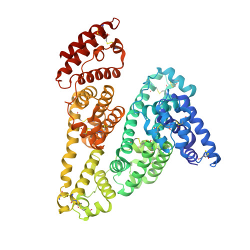
Glucose reacts with proteins nonenzymatically under physiological conditions. Such glycation is exacerbated in diabetic patients with high levels of blood sugar and induces various complications. Human albumin serum (HSA) is the most abundant protein in plasma and is glycated by glucose. The glycation sites on HSA remain controversial among different studies. Here, we report two protein crystal structures of HSA in complex with either glucose or fructose. These crystal structures reveal the presence of linear forms of sugar for both monosaccharides. The linear form of glucose forms a covalent bond to Lys-195 of HSA, but this is not the case for fructose. Based on these structures, we propose a mechanism for glucose ring opening involving both residues Lys-195 and Lys-199. These results provide mechanistic insights to understand the glucose ring-opening reaction and the glycation of proteins by monosaccharides.
- State Key Laboratory of Structural Chemistry, Fujian Institute of Research on the Structure of Matter, Chinese Academy of Sciences, Fuzhou, Fujian 350002, China.
Organizational Affiliation: