4IM9 | pdb_00004im9

Cystal structure of DnaG primase C-terminal domain from Vibrio cholerae


Experimental Data Snapshot

  • Method: X-RAY DIFFRACTION
  • Resolution: 2.46 Å
  • R-Value Free: 
    0.264 (Depositor), 0.262 (DCC) 
  • R-Value Work: 
    0.227 (Depositor), 0.231 (DCC) 
  • R-Value Observed: 
    0.229 (Depositor) 

wwPDB Validation   3D Report Full Report


This is version 1.1 of the entry. See complete history


Literature

Cystal structure of DnaG primase C-terminal domain from Vibrio cholerae

Abdul Rehman, S.A.Tarique, K.F.Gourinath, S.

To be published.

Macromolecules
Find similar proteins by:  (by identity cutoff)  |  3D Structure
Entity ID: 1
MoleculeChains Sequence LengthOrganismDetailsImage
DNA primase
A, B, C
152Vibrio cholerae O395Mutation(s): 0 
Gene Names: C-terminal domaindnaGPrimaseVC395_0535
EC: 2.7.7
Entity Groups  
Sequence Clusters30% Identity50% Identity70% Identity90% Identity95% Identity100% Identity
Sequence Annotations
Expand
  • Reference Sequence
Small Molecules
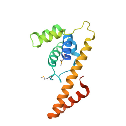
Modified Residues  1 Unique
IDChains TypeFormula2D DiagramParent
MSE
Query on MSE
A, B, C
L-PEPTIDE LINKINGC5 H11 N O2 SeMET
Experimental Data & Validation

Experimental Data

  • Method: X-RAY DIFFRACTION
  • Resolution: 2.46 Å
  • R-Value Free:  0.264 (Depositor), 0.262 (DCC) 
  • R-Value Work:  0.227 (Depositor), 0.231 (DCC) 
  • R-Value Observed: 0.229 (Depositor) 
Space Group: P 21 21 21
Unit Cell:
Length ( Å )Angle ( ˚ )
a = 68.412α = 90
b = 73.779β = 90
c = 132.653γ = 90
Software Package:
Software NamePurpose
DNAdata collection
SHELXSphasing
REFMACrefinement
HKL-2000data reduction
HKL-2000data scaling

Structure Validation

View Full Validation Report


Entry History 

Deposition Data

Revision History  (Full details and data files)

  • Version 1.0: 2014-05-07
    Type: Initial release
  • Version 1.1: 2024-11-20
    Changes: Data collection, Database references, Derived calculations, Structure summary