4I6L

Crystal structure of OTUB1 in complex with ubiquitin variant


Experimental Data Snapshot

  • Method: X-RAY DIFFRACTION
  • Resolution: 2.49 Å
  • R-Value Free: 0.266 
  • R-Value Work: 0.220 
  • R-Value Observed: 0.222 

wwPDB Validation   3D Report Full Report


This is version 1.3 of the entry. See complete history


Literature

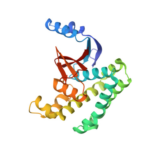
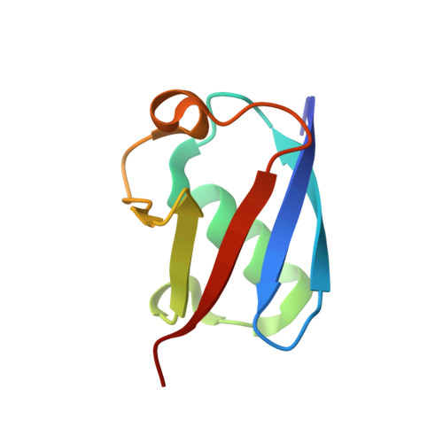
A strategy for modulation of enzymes in the ubiquitin system.

Ernst, A.Avvakumov, G.Tong, J.Fan, Y.Zhao, Y.Alberts, P.Persaud, A.Walker, J.R.Neculai, A.M.Neculai, D.Vorobyov, A.Garg, P.Beatty, L.Chan, P.K.Juang, Y.C.Landry, M.C.Yeh, C.Zeqiraj, E.Karamboulas, K.Allali-Hassani, A.Vedadi, M.Tyers, M.Moffat, J.Sicheri, F.Pelletier, L.Durocher, D.Raught, B.Rotin, D.Yang, J.Moran, M.F.Dhe-Paganon, S.Sidhu, S.S.

(2013) Science 339: 590-595

  • DOI: https://doi.org/10.1126/science.1230161
  • Primary Citation of Related Structures:  
    3I3T, 3MTN, 3N3K, 3V6E, 4I6L

  • PubMed Abstract: 

    The ubiquitin system regulates virtually all aspects of cellular function. We report a method to target the myriad enzymes that govern ubiquitination of protein substrates. We used massively diverse combinatorial libraries of ubiquitin variants to develop inhibitors of four deubiquitinases (DUBs) and analyzed the DUB-inhibitor complexes with crystallography. We extended the selection strategy to the ubiquitin conjugating (E2) and ubiquitin ligase (E3) enzymes and found that ubiquitin variants can also enhance enzyme activity. Last, we showed that ubiquitin variants can bind selectively to ubiquitin-binding domains. Ubiquitin variants exhibit selective function in cells and thus enable orthogonal modulation of specific enzymatic steps in the ubiquitin system.


  • Organizational Affiliation

    Terrence Donnelly Center for Cellular and Biomolecular Research, University of Toronto, 160 College Street, Toronto, Ontario, Canada.


Macromolecules
Find similar proteins by:  (by identity cutoff)  |  3D Structure
Entity ID: 1
MoleculeChains Sequence LengthOrganismDetailsImage
Ubiquitin thioesterase OTUB1229Homo sapiensMutation(s): 1 
Gene Names: OTUB1OTB1OTU1HSPC263
EC: 3.4.19.12
UniProt & NIH Common Fund Data Resources
Find proteins for Q96FW1 (Homo sapiens)
Explore Q96FW1 
Go to UniProtKB:  Q96FW1
PHAROS:  Q96FW1
GTEx:  ENSG00000167770 
Entity Groups  
Sequence Clusters30% Identity50% Identity70% Identity90% Identity95% Identity100% Identity
UniProt GroupQ96FW1
Sequence Annotations
Expand
  • Reference Sequence
Find similar proteins by:  (by identity cutoff)  |  3D Structure
Entity ID: 2
MoleculeChains Sequence LengthOrganismDetailsImage
Ubiquitin76Homo sapiensMutation(s): 6 
Gene Names: UBC
UniProt & NIH Common Fund Data Resources
Find proteins for P0CG48 (Homo sapiens)
Explore P0CG48 
Go to UniProtKB:  P0CG48
PHAROS:  P0CG48
GTEx:  ENSG00000150991 
Entity Groups  
Sequence Clusters30% Identity50% Identity70% Identity90% Identity95% Identity100% Identity
UniProt GroupP0CG48
Sequence Annotations
Expand
  • Reference Sequence
Experimental Data & Validation

Experimental Data

  • Method: X-RAY DIFFRACTION
  • Resolution: 2.49 Å
  • R-Value Free: 0.266 
  • R-Value Work: 0.220 
  • R-Value Observed: 0.222 
  • Space Group: C 2 2 21
Unit Cell:
Length ( Å )Angle ( ˚ )
a = 92.444α = 90
b = 109.069β = 90
c = 73.238γ = 90
Software Package:
Software NamePurpose
HKL-2000data collection
PHASERphasing
PHENIXrefinement
HKL-2000data reduction
HKL-2000data scaling

Structure Validation

View Full Validation Report


Entry History 

Deposition Data

Revision History  (Full details and data files)

  • Version 1.0: 2013-01-16
    Type: Initial release
  • Version 1.1: 2013-02-06
    Changes: Database references
  • Version 1.2: 2013-02-20
    Changes: Database references
  • Version 1.3: 2023-09-20
    Changes: Data collection, Database references, Refinement description