


Propionaldehyde does not bind to PduB
Pang, A.H., Prentice, M.B., Pickersgill, R.W.To be published.
Experimental Data Snapshot
Starting Model: experimental
View more details
wwPDB Validation 3D Report Full Report

Entity ID: 1 | |||||
|---|---|---|---|---|---|
| Molecule | Chains | Sequence Length | Organism | Details | Image |
| Propanediol utilization protein PduB | 258 | Limosilactobacillus reuteri SD2112 | Mutation(s): 0 Gene Names: HMPREF0538_20934, pduB | 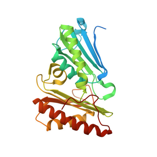 | |
UniProt | |||||
Entity Groups | |||||
| Sequence Clusters | 30% Identity50% Identity70% Identity90% Identity95% Identity100% Identity | ||||
| UniProt Group | F8DQ39 | ||||
Sequence AnnotationsExpand | |||||
Reference Sequence | |||||
| Length ( Å ) | Angle ( ˚ ) |
|---|---|
| a = 69.01 | α = 90 |
| b = 119.55 | β = 90 |
| c = 147.09 | γ = 90 |
| Software Name | Purpose |
|---|---|
| ADSC | data collection |
| PHENIX | model building |
| REFMAC | refinement |
| XDS | data reduction |
| SCALA | data scaling |
| PHENIX | phasing |


