4I49 | pdb_00004i49

Structure of ngNAGS bound with bisubstrate analog CoA-NAG


Experimental Data Snapshot

  • Method: X-RAY DIFFRACTION
  • Resolution: 2.75 Å
  • R-Value Free: 
    0.271 (Depositor), 0.264 (DCC) 
  • R-Value Work: 
    0.189 (Depositor), 0.184 (DCC) 
  • R-Value Observed: 
    0.198 (Depositor) 

Starting Model: experimental
View more details

wwPDB Validation 3D Report Full Report

Validation slider image for 4I49

Ligand Structure Quality Assessment 


This is version 1.3 of the entry. See complete history

Literature

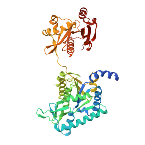
Structure of the complex of Neisseria gonorrhoeae N-acetyl-l-glutamate synthase with a bound bisubstrate analog.

Zhao, G.Allewell, N.M.Tuchman, M.Shi, D.

(2013) Biochem Biophys Res Commun 430: 1253-1258

  • DOI: https://doi.org/10.1016/j.bbrc.2012.12.064
  • Primary Citation Related Structures: 
    4I49

  • PubMed Abstract: 

    N-Acetyl-L-glutamate synthase catalyzes the conversion of AcCoA and glutamate to CoA and N-acetyl-L-glutamate (NAG), the first step of the arginine biosynthetic pathway in lower organisms. In mammals, NAG is an obligate cofactor of carbamoyl phosphate synthetase I in the urea cycle. We have previously reported the structures of NAGS from Neisseria gonorrhoeae (ngNAGS) with various substrates bound. Here we reported the preparation of the bisubstrate analog, CoA-S-acetyl-L-glutamate, the crystal structure of ngNAGS with CoA-NAG bound, and kinetic studies of several active site mutants. The results are consistent with a one-step nucleophilic addition-elimination mechanism with Glu353 as the catalytic base and Ser392 as the catalytic acid. The structure of the ngNAGS-bisubstrate complex together with the previous ngNAGS structures delineates the catalytic reaction path for ngNAGS.


  • Organizational Affiliation
    • Center for Genetic Medicine Research and Department of Integrative Systems Biology, Children's National Medical Center, The George Washington University, Washington, DC 20010, USA.

Macromolecule Content 

  • Total Structure Weight: 50.29 kDa 
  • Atom Count: 3,327 
  • Modeled Residue Count: 424 
  • Deposited Residue Count: 456 
  • Unique protein chains: 1

Macromolecules

Find similar proteins by:|  3D Structure
Entity ID: 1
MoleculeChains  Sequence LengthOrganismDetailsImage
Amino-acid acetyltransferase456Neisseria gonorrhoeae SK-93-1035Mutation(s): 0 
Gene Names: argANGLG_00316
EC: 2.3.1.1

Small Molecules

Ligands 2 Unique
IDChains Name / Formula / InChI Key2D Diagram3D Interactions
1C4

Query on 1C4


Download:Ideal Coordinates CCD File
B [auth A](2S)-2-({(3S,5R,9R)-1-[(2R,3S,4R,5R)-5-(6-amino-9H-purin-9-yl)-4-hydroxy-3-(phosphonooxy)tetrahydrofuran-2-yl]-3,5,9-trihydroxy-8,8-dimethyl-3,5-dioxido-10,14,20-trioxo-2,4,6-trioxa-18-thia-11,15-diaza-3lambda~5~,5lambda~5~-diphosphaicosan-20-yl}amino)pentanedioic acid (non-preferred name)
C28 H45 N8 O21 P3 S
WCWCYRFADJEDNS-NOTSHUFBSA-N
SO4

Query on SO4


Download:Ideal Coordinates CCD File
C [auth A]SULFATE ION
O4 S
QAOWNCQODCNURD-UHFFFAOYSA-L

Experimental Data & Validation

Experimental Data

  • Method: X-RAY DIFFRACTION
  • Resolution: 2.75 Å
  • R-Value Free:  0.271 (Depositor), 0.264 (DCC) 
  • R-Value Work:  0.189 (Depositor), 0.184 (DCC) 
  • R-Value Observed: 0.198 (Depositor) 
Space Group: P 3 1 2
Unit Cell:
Length ( Å )Angle ( ˚ )
a = 98.521α = 90
b = 98.521β = 90
c = 90.009γ = 120
Software Package:
Software NamePurpose
DENZOdata reduction
SCALEPACKdata scaling
PHENIXrefinement
PDB_EXTRACTdata extraction
StructureStudiodata collection

Structure Validation

View Full Validation Report


Ligand Structure Quality Assessment 


Entry History 

Deposition Data

Revision History  (Full details and data files)

  • Version 1.0: 2013-01-09
    Type: Initial release
  • Version 1.1: 2013-03-06
    Changes: Database references
  • Version 1.2: 2017-11-15
    Changes: Refinement description
  • Version 1.3: 2023-09-20
    Changes: Data collection, Database references, Derived calculations, Refinement description