4HVP | pdb_00004hvp

Structure of complex of synthetic HIV-1 protease with a substrate-based inhibitor at 2.3 Angstroms resolution


Experimental Data Snapshot

  • Method: X-RAY DIFFRACTION
  • Resolution: 2.30 Å
  • R-Value Observed: 
    0.176 (Depositor) 

wwPDB Validation 3D Report Full Report

Validation slider image for 4HVP

This is version 1.6 of the entry. See complete history

Literature

Structure of complex of synthetic HIV-1 protease with a substrate-based inhibitor at 2.3 A resolution.

Miller, M.Schneider, J.Sathyanarayana, B.K.Toth, M.V.Marshall, G.R.Clawson, L.Selk, L.Kent, S.B.Wlodawer, A.

(1989) Science 246: 1149-1152

  • DOI: https://doi.org/10.1126/science.2686029
  • Primary Citation Related Structures: 
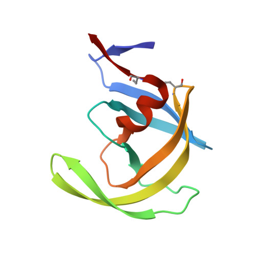
    4HVP

  • PubMed Abstract: 

    The structure of a complex between a peptide inhibitor with the sequence N-acetyl-Thr-Ile-Nle-psi[CH2-NH]-Nle-Gln-Arg.amide (Nle, norleucine) with chemically synthesized HIV-1 (human immunodeficiency virus 1) protease was determined at 2.3 A resolution (R factor of 0.176). Despite the symmetric nature of the unliganded enzyme, the asymmetric inhibitor lies in a single orientation and makes extensive interactions at the interface between the two subunits of the homodimeric protein. Compared with the unliganded enzyme, the protein molecule underwent substantial changes, particularly in an extended region corresponding to the "flaps" (residues 35 to 57 in each chain), where backbone movements as large as 7 A are observed.


  • Organizational Affiliation
    • NCI-Frederick Cancer Research Facility, BRI-Basic Research Program, MD 21701.

Macromolecule Content 

  • Total Structure Weight: 22.3 kDa 
  • Atom Count: 1,640 
  • Modeled Residue Count: 198 
  • Deposited Residue Count: 198 
  • Unique protein chains: 1

Macromolecules

Find similar proteins by:|  3D Structure
Entity ID: 1
MoleculeChains  Sequence LengthOrganismDetailsImage
HIV-1 PROTEASE
A, B
99Human immunodeficiency virus 1Mutation(s): 0 
UniProt
Find proteins for P03369 (Human immunodeficiency virus type 1 group M subtype B (isolate ARV2/SF2))
Explore P03369 
Go to UniProtKB:  P03369
Entity Groups
Sequence Clusters30% Identity50% Identity70% Identity90% Identity95% Identity100% Identity
UniProt GroupP03369
Sequence Annotations
Expand
Reference Sequence

Small Molecules

Ligands 1 Unique
IDChains Name / Formula / InChI Key2D Diagram3D Interactions
2NC

Query on 2NC


Download:Ideal Coordinates CCD File
C [auth B]N-{(2S)-2-[(N-acetyl-L-threonyl-L-isoleucyl)amino]hexyl}-L-norleucyl-L-glutaminyl-N~5~-[amino(iminio)methyl]-L-ornithinamide
C35 H68 N11 O8
MQPXOVRKKPPKFZ-QYKDHROSSA-O
Modified Residues  1 Unique
IDChains TypeFormula2D DiagramParent
ABA
Query on ABA
A, B
L-PEPTIDE LINKINGC4 H9 N O2ALA

Biologically Interesting Molecules (External Reference) 

1 Unique

Experimental Data & Validation

Experimental Data

  • Method: X-RAY DIFFRACTION
  • Resolution: 2.30 Å
  • R-Value Observed: 0.176 (Depositor) 
Space Group: P 21 21 21
Unit Cell:
Length ( Å )Angle ( ˚ )
a = 51.7α = 90
b = 59.2β = 90
c = 62.45γ = 90
Software Package:
Software NamePurpose
PROLSQrefinement

Structure Validation

View Full Validation Report


Entry History 

Deposition Data

Revision History  (Full details and data files)

  • Version 1.0: 1990-04-15
    Type: Initial release
  • Version 1.1: 2008-03-03
    Changes: Version format compliance
  • Version 1.2: 2011-07-13
    Changes: Atomic model, Database references, Derived calculations, Non-polymer description, Structure summary, Version format compliance
  • Version 1.3: 2013-02-27
    Changes: Other
  • Version 1.4: 2013-04-03
    Changes: Structure summary
  • Version 1.5: 2017-11-29
    Changes: Derived calculations, Other
  • Version 1.6: 2025-03-26
    Changes: Data collection, Database references, Derived calculations, Structure summary