4HM8

Naphthalene 1,2-Dioxygenase bound to thioanisole


Experimental Data Snapshot

  • Method: X-RAY DIFFRACTION
  • Resolution: 1.30 Å
  • R-Value Free: 0.194 
  • R-Value Work: 0.170 
  • R-Value Observed: 0.171 

wwPDB Validation   3D Report Full Report


This is version 1.2 of the entry. See complete history


Literature

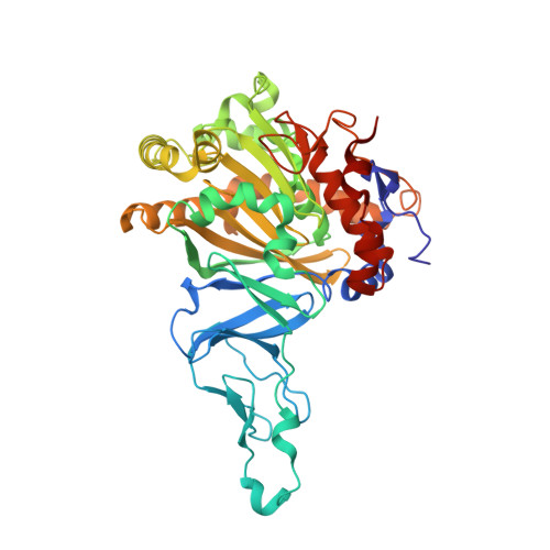
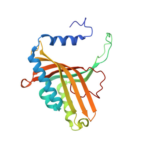
One enzyme, many reactions: structural basis for the various reactions catalyzed by naphthalene 1,2-dioxygenase.

Ferraro, D.J.Okerlund, A.Brown, E.Ramaswamy, S.

(2017) IUCrJ 4: 648-656

  • DOI: https://doi.org/10.1107/S2052252517008223
  • Primary Citation of Related Structures:  
    4HJL, 4HKV, 4HM0, 4HM2, 4HM3, 4HM4, 4HM5, 4HM6, 4HM7, 4HM8

  • PubMed Abstract: 

    Rieske nonheme iron oxygenases (ROs) are a well studied class of enzymes. Naphthalene 1,2-dioxygenase (NDO) is used as a model to study ROs. Previous work has shown how side-on binding of oxygen to the mononuclear iron provides this enzyme with the ability to catalyze stereospecific and regiospecific cis -dihydroxylation reactions. It has been well documented that ROs catalyze a variety of other reactions, including mono-oxygenation, desaturation, O- and N-dealkylation, sulfoxidation etc . NDO itself catalyzes a variety of these reactions. Structures of NDO in complex with a number of different substrates show that the orientation of the substrate in the active site controls not only the regiospecificity and stereospecificity, but also the type of reaction catalyzed. It is proposed that the mononuclear iron-activated dioxygen attacks the atoms of the substrate that are most proximal to it. The promiscuity of delivering two products (apparently by two different reactions) from the same substrate can be explained by the possible binding of the substrate in slightly different orientations aided by the observed flexibility of residues in the binding pocket.


  • Organizational Affiliation

    Department of Biochemistry, Carver College of Medicine, University of Iowa, Iowa City, IA 52242, USA.


Macromolecules
Find similar proteins by:  (by identity cutoff)  |  3D Structure
Entity ID: 1
MoleculeChains Sequence LengthOrganismDetailsImage
Naphthalene 1,2-dioxygenase subunit alpha449Pseudomonas sp. C18Mutation(s): 0 
Gene Names: doxBNC_004999.1
EC: 1.14.12.12
UniProt
Find proteins for P0A111 (Pseudomonas sp. (strain C18))
Explore P0A111 
Go to UniProtKB:  P0A111
Entity Groups  
Sequence Clusters30% Identity50% Identity70% Identity90% Identity95% Identity100% Identity
UniProt GroupP0A111
Sequence Annotations
Expand
  • Reference Sequence
Find similar proteins by:  (by identity cutoff)  |  3D Structure
Entity ID: 2
MoleculeChains Sequence LengthOrganismDetailsImage
Naphthalene 1,2-dioxygenase subunit beta194Pseudomonas sp. C18Mutation(s): 0 
Gene Names: doxDNC_004999.1
EC: 1.14.12.12
UniProt
Find proteins for P0A113 (Pseudomonas sp. (strain C18))
Explore P0A113 
Go to UniProtKB:  P0A113
Entity Groups  
Sequence Clusters30% Identity50% Identity70% Identity90% Identity95% Identity100% Identity
UniProt GroupP0A113
Sequence Annotations
Expand
  • Reference Sequence
Small Molecules
Ligands 5 Unique
IDChains Name / Formula / InChI Key2D Diagram3D Interactions
FES
Query on FES

Download Ideal Coordinates CCD File 
F [auth A]FE2/S2 (INORGANIC) CLUSTER
Fe2 S2
NIXDOXVAJZFRNF-UHFFFAOYSA-N
16R
Query on 16R

Download Ideal Coordinates CCD File 
H [auth A](methylsulfanyl)benzene
C7 H8 S
HNKJADCVZUBCPG-UHFFFAOYSA-N
SO4
Query on SO4

Download Ideal Coordinates CCD File 
E [auth A],
K [auth A],
O [auth B],
P [auth B]
SULFATE ION
O4 S
QAOWNCQODCNURD-UHFFFAOYSA-L
EDO
Query on EDO

Download Ideal Coordinates CCD File 
C [auth A]
D [auth A]
I [auth A]
J [auth A]
L [auth B]
C [auth A],
D [auth A],
I [auth A],
J [auth A],
L [auth B],
M [auth B],
N [auth B],
Q [auth B]
1,2-ETHANEDIOL
C2 H6 O2
LYCAIKOWRPUZTN-UHFFFAOYSA-N
FE
Query on FE

Download Ideal Coordinates CCD File 
G [auth A]FE (III) ION
Fe
VTLYFUHAOXGGBS-UHFFFAOYSA-N
Experimental Data & Validation

Experimental Data

  • Method: X-RAY DIFFRACTION
  • Resolution: 1.30 Å
  • R-Value Free: 0.194 
  • R-Value Work: 0.170 
  • R-Value Observed: 0.171 
  • Space Group: H 3 2
Unit Cell:
Length ( Å )Angle ( ˚ )
a = 140.236α = 90
b = 140.236β = 90
c = 208.29γ = 120
Software Package:
Software NamePurpose
d*TREKdata reduction
PHENIXrefinement
PDB_EXTRACTdata extraction
d*TREKdata scaling
AMoREphasing

Structure Validation

View Full Validation Report


Entry History 

Deposition Data

Revision History  (Full details and data files)

  • Version 1.0: 2013-10-30
    Type: Initial release
  • Version 1.1: 2016-04-06
    Changes: Non-polymer description
  • Version 1.2: 2024-09-18
    Changes: Data collection, Database references, Derived calculations