4HB1 | pdb_00004hb1

A DESIGNED FOUR HELIX BUNDLE PROTEIN.


Experimental Data Snapshot

  • Method: X-RAY DIFFRACTION
  • Resolution: 2.90 Å
  • R-Value Free: 
    0.284 (Depositor), 0.332 (DCC) 
  • R-Value Work: 
    0.234 (Depositor), 0.251 (DCC) 
  • R-Value Observed: 
    0.234 (Depositor) 

Starting Model: in silico
View more details

wwPDB Validation 3D Report Full Report

Validation slider image for 4HB1

This is version 1.4 of the entry. See complete history

Literature

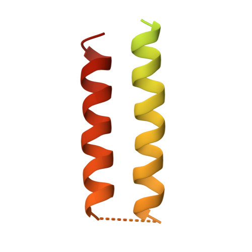
A designed four helix bundle protein with native-like structure.

Schafmeister, C.E.LaPorte, S.L.Miercke, L.J.Stroud, R.M.

(1997) Nat Struct Biol 4: 1039-1046

  • DOI: https://doi.org/10.1038/nsb1297-1039
  • Primary Citation Related Structures: 
    4HB1

  • PubMed Abstract: 

    A 108 amino acid protein was designed and constructed from a reduced alphabet of seven amino acids. The 2.9 A resolution X-ray crystal structure confirms that the protein is a four helix bundle, as it was designed to be. Hydrogen/deuterium exchange experiments reveal buried amide protons with protection factors in excess of 1 x 10(6) in the range characteristic of well protected protons in functional folded proteins (10(3)-10(8)) rather than protons in rapid exchange (0-10(2)). The protein is monomeric at 1 mM, the concentration at which the exchange experiments were undertaken, indicating that the exchange factors are due to a unique stable tertiary structure fold, and not due to any higher order quaternary structure. Thermodynamic analysis provides an estimate of the free energy of folding of -9.3 kcal mole-1 at 25 degrees C, consistent with the free energy of folding derived from the protection factors of the most protected protons, indicating that global unfolding is required for exchange of the most protected protons.


  • Organizational Affiliation
    • Department of Biochemistry and Biophysics, University of California, San Francisco 94143-0448, USA.

Macromolecule Content 

  • Total Structure Weight: 11.82 kDa 
  • Atom Count: 300 
  • Modeled Residue Count: 44 
  • Deposited Residue Count: 108 
  • Unique protein chains: 1

Macromolecules

Find similar proteins by:|  3D Structure
Entity ID: 1
MoleculeChains  Sequence LengthOrganismDetailsImage
DHP1108synthetic constructMutation(s): 0 
Gene Names: SYNTHETIC GENE DHP1

Experimental Data & Validation

Experimental Data

  • Method: X-RAY DIFFRACTION
  • Resolution: 2.90 Å
  • R-Value Free:  0.284 (Depositor), 0.332 (DCC) 
  • R-Value Work:  0.234 (Depositor), 0.251 (DCC) 
  • R-Value Observed: 0.234 (Depositor) 
Space Group: P 64 2 2
Unit Cell:
Length ( Å )Angle ( ˚ )
a = 51.9α = 90
b = 51.9β = 90
c = 61γ = 120
Software Package:
Software NamePurpose
X-PLORmodel building
X-PLORrefinement
DENZOdata reduction
X-PLORphasing

Structure Validation

View Full Validation Report


Entry History 

Deposition Data

Revision History  (Full details and data files)

  • Version 1.0: 1998-03-04
    Type: Initial release
  • Version 1.1: 2008-03-25
    Changes: Version format compliance
  • Version 1.2: 2011-07-13
    Changes: Version format compliance
  • Version 1.3: 2024-02-28
    Changes: Data collection, Database references, Derived calculations, Other
  • Version 1.4: 2024-04-03
    Changes: Refinement description