4H8I | pdb_00004h8i

Structure of GluK2-LBD in complex with GluAzo


Experimental Data Snapshot

  • Method: X-RAY DIFFRACTION
  • Resolution: 2.00 Å
  • R-Value Free: 
    0.204 (Depositor), 0.202 (DCC) 
  • R-Value Work: 
    0.173 (Depositor), 0.172 (DCC) 
  • R-Value Observed: 
    0.175 (Depositor) 

Starting Model: experimental
View more details

wwPDB Validation 3D Report Full Report

Validation slider image for 4H8I

Ligand Structure Quality Assessment 


This is version 1.5 of the entry. See complete history

Literature

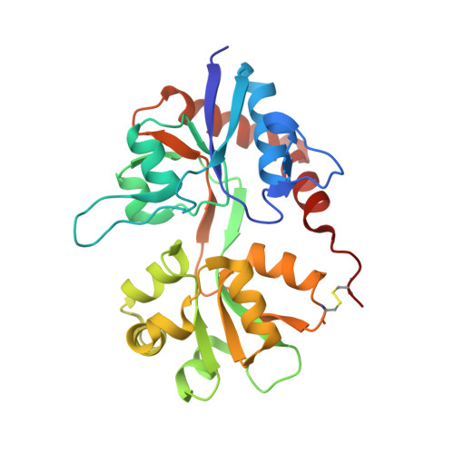
A photoswitchable neurotransmitter analogue bound to its receptor.

Reiter, A.Skerra, A.Trauner, D.Schiefner, A.

(2013) Biochemistry 52: 8972-8974

  • DOI: https://doi.org/10.1021/bi4014402
  • Primary Citation Related Structures: 
    4H8I

  • PubMed Abstract: 

    Incorporation of the azobenzene derivative gluazo, a synthetic photochromic ligand, into a kainate receptor allows for the optical control of neuronal activity. The crystal structure of gluazo bound to a dimeric GluK2 ligand-binding domain reveals one monomer in a closed conformation, occupied by gluazo, and the other in an open conformation, with a bound buffer molecule. The glutamate group of gluazo interacts like the natural glutamate ligand, while its trans-azobenzene moiety protrudes into a tunnel. This elongated cavity presumably cannot accommodate a cis-azobenzene, which explains the reversible activation of the receptor upon photoisomerization.


  • Organizational Affiliation
    • Munich Center for Integrated Protein Science (CIPS-M).

Macromolecule Content 

  • Total Structure Weight: 59.44 kDa 
  • Atom Count: 4,645 
  • Modeled Residue Count: 512 
  • Deposited Residue Count: 518 
  • Unique protein chains: 1

Macromolecules

Find similar proteins by:|  3D Structure
Entity ID: 1
MoleculeChains  Sequence LengthOrganismDetailsImage
Glutamate receptor, ionotropic kainate 2
A, B
259Rattus norvegicusMutation(s): 0 
Gene Names: Glur6Grik2
UniProt
Find proteins for P42260 (Rattus norvegicus)
Explore P42260 
Go to UniProtKB:  P42260
Entity Groups
Sequence Clusters30% Identity50% Identity70% Identity90% Identity95% Identity100% Identity
UniProt GroupP42260
Sequence Annotations
Expand
Reference Sequence

Experimental Data & Validation

Experimental Data

  • Method: X-RAY DIFFRACTION
  • Resolution: 2.00 Å
  • R-Value Free:  0.204 (Depositor), 0.202 (DCC) 
  • R-Value Work:  0.173 (Depositor), 0.172 (DCC) 
  • R-Value Observed: 0.175 (Depositor) 
Space Group: P 61 2 2
Unit Cell:
Length ( Å )Angle ( ˚ )
a = 102.609α = 90
b = 102.609β = 90
c = 282.513γ = 120
Software Package:
Software NamePurpose
XSCALEdata scaling
PHASERphasing
REFMACrefinement
PDB_EXTRACTdata extraction
MAR345data collection
XDSdata reduction

Structure Validation

View Full Validation Report


Ligand Structure Quality Assessment 


Entry History 

Deposition Data

Revision History  (Full details and data files)

  • Version 1.0: 2013-09-25
    Type: Initial release
  • Version 1.1: 2014-12-24
    Changes: Database references
  • Version 1.2: 2017-07-26
    Changes: Refinement description, Source and taxonomy
  • Version 1.3: 2017-11-15
    Changes: Refinement description
  • Version 1.4: 2023-09-20
    Changes: Data collection, Database references, Derived calculations, Refinement description
  • Version 1.5: 2024-11-27
    Changes: Structure summary