4H5T

HSC70 NBD with ADP and Mg


Experimental Data Snapshot

  • Method: X-RAY DIFFRACTION
  • Resolution: 1.90 Å
  • R-Value Free: 0.298 
  • R-Value Work: 0.232 
  • R-Value Observed: 0.235 

wwPDB Validation   3D Report Full Report


Ligand Structure Quality Assessment 


This is version 1.3 of the entry. See complete history


Literature

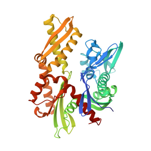
New crystal structures of HSC-70 ATP binding domain confirm the role of individual binding pockets and suggest a new method of inhibition.

Zhang, Z.Cellitti, J.Teriete, P.Pellecchia, M.Stec, B.

(2015) Biochimie 108: 186-192

  • DOI: https://doi.org/10.1016/j.biochi.2014.11.012
  • Primary Citation of Related Structures:  
    4H5N, 4H5R, 4H5T, 4H5V, 4H5W

  • PubMed Abstract: 

    In recent years the chaperone HSC-70 has become a target for drug design with a strong focus in anticancer therapies. In our study of possible inhibitors of HSC-70 enzymatic activity we screened compounds by NMR as well as X-ray crystallography. As part of our screening efforts we crystallized the human HSC-70 ATP binding domain and obtained novel crystal forms in addition to known structures. The new crystal structures highlight the mobility of the entire domain previously described by NMR, which was linked to its chaperone activity but not yet demonstrated by X-ray crystallography. Conformational changes across the entire molecule have been elucidated in response to the binding of small molecule ligands and show a pattern of mobility consistent with postulated signal transduction modes between the nucleotide binding domain (NBD) and the substrate binding domain (SBD). In addition, two crystal structures contained glycerol bound at a new site. Binding studies performed with glycerol analogs proved inhibitory properties of the site, which were further characterized by isothermal calorimetry and in silico docking studies. The presence of two binding pockets enabled us to explore a novel method of inhibition by compounds that bridge the adjacent phosphate and glycerol binding sites. Finally, an example of such a bridged inhibitor is proposed.


  • Organizational Affiliation

    Sanford-Burnham Medical Research Institute, 10901 N. Torrey Pines Rd., La Jolla, CA 92037, USA.


Macromolecules
Find similar proteins by:  (by identity cutoff)  |  3D Structure
Entity ID: 1
MoleculeChains Sequence LengthOrganismDetailsImage
Heat shock cognate 71 kDa protein383Homo sapiensMutation(s): 0 
Gene Names: HSC70HSP73HSPA10HSPA8
UniProt & NIH Common Fund Data Resources
Find proteins for P11142 (Homo sapiens)
Explore P11142 
Go to UniProtKB:  P11142
PHAROS:  P11142
GTEx:  ENSG00000109971 
Entity Groups  
Sequence Clusters30% Identity50% Identity70% Identity90% Identity95% Identity100% Identity
UniProt GroupP11142
Sequence Annotations
Expand
  • Reference Sequence
Small Molecules
Binding Affinity Annotations 
IDSourceBinding Affinity
ADP BindingDB:  4H5T Kd: min: 320, max: 324 (nM) from 2 assay(s)
IC50: 280 (nM) from 1 assay(s)
Experimental Data & Validation

Experimental Data

  • Method: X-RAY DIFFRACTION
  • Resolution: 1.90 Å
  • R-Value Free: 0.298 
  • R-Value Work: 0.232 
  • R-Value Observed: 0.235 
  • Space Group: C 1 2 1
Unit Cell:
Length ( Å )Angle ( ˚ )
a = 131.905α = 90
b = 46.379β = 127.2
c = 87.711γ = 90
Software Package:
Software NamePurpose
CrystalCleardata collection
MOLREPphasing
REFMACrefinement
HKL-2000data reduction
SCALEPACKdata scaling

Structure Validation

View Full Validation Report


Ligand Structure Quality Assessment 


Entry History 

Deposition Data

  • Released Date: 2014-03-19 
  • Deposition Author(s): Stec, B.

Revision History  (Full details and data files)

  • Version 1.0: 2014-03-19
    Type: Initial release
  • Version 1.1: 2014-12-17
    Changes: Database references
  • Version 1.2: 2015-01-14
    Changes: Database references
  • Version 1.3: 2023-09-20
    Changes: Data collection, Database references, Derived calculations, Refinement description