4GZ6

Crystal structure of human O-GlcNAc Transferase with UDP-5SGlcNAc


Experimental Data Snapshot

  • Method: X-RAY DIFFRACTION
  • Resolution: 2.98 Å
  • R-Value Free: 0.223 
  • R-Value Work: 0.197 
  • R-Value Observed: 0.198 

wwPDB Validation   3D Report Full Report


Ligand Structure Quality Assessment 


This is version 1.2 of the entry. See complete history


Literature

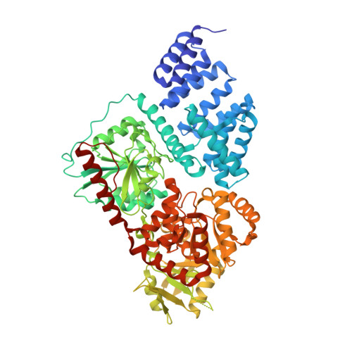
Structural snapshots of the reaction coordinate for O-GlcNAc transferase.

Lazarus, M.B.Jiang, J.Gloster, T.M.Zandberg, W.F.Whitworth, G.E.Vocadlo, D.J.Walker, S.

(2012) Nat Chem Biol 8: 966-968

  • DOI: https://doi.org/10.1038/nchembio.1109
  • Primary Citation of Related Structures:  
    4GYY, 4GZ3, 4GZ5, 4GZ6

  • PubMed Abstract: 

    Visualization of the reaction coordinate undertaken by glycosyltransferases has remained elusive but is critical for understanding this important class of enzyme. Using substrates and substrate mimics, we describe structural snapshots of all species along the kinetic pathway for human O-linked β-N-acetylglucosamine transferase (O-GlcNAc transferase), an intracellular enzyme that catalyzes installation of a dynamic post-translational modification. The structures reveal key features of the mechanism and show that substrate participation is important during catalysis.


  • Organizational Affiliation

    Department of Chemistry and Chemical Biology, Harvard University, Cambridge, Massachusetts, USA.


Macromolecules
Find similar proteins by:  (by identity cutoff)  |  3D Structure
Entity ID: 1
MoleculeChains Sequence LengthOrganismDetailsImage
UDP-N-acetylglucosamine--peptide N-acetylglucosaminyltransferase 110 kDa subunit
A, B, C, D
723Homo sapiensMutation(s): 0 
Gene Names: OGT
EC: 2.4.1.255
UniProt & NIH Common Fund Data Resources
Find proteins for O15294 (Homo sapiens)
Explore O15294 
Go to UniProtKB:  O15294
PHAROS:  O15294
GTEx:  ENSG00000147162 
Entity Groups  
Sequence Clusters30% Identity50% Identity70% Identity90% Identity95% Identity100% Identity
UniProt GroupO15294
Sequence Annotations
Expand
  • Reference Sequence
Small Molecules
Ligands 2 Unique
IDChains Name / Formula / InChI Key2D Diagram3D Interactions
12V
Query on 12V

Download Ideal Coordinates CCD File 
E [auth A],
J [auth B],
P [auth C],
U [auth D]
(2S,3R,4R,5S,6R)-3-(acetylamino)-4,5-dihydroxy-6-(hydroxymethyl)tetrahydro-2H-thiopyran-2-yl [(2R,3S,4R,5R)-5-(2,4-dioxo-3,4-dihydropyrimidin-1(2H)-yl)-3,4-dihydroxytetrahydrofuran-2-yl]methyl dihydrogen diphosphate
C17 H27 N3 O16 P2 S
JPRVHSQHWXZSNC-UBDZBXRQSA-N
SO4
Query on SO4

Download Ideal Coordinates CCD File 
F [auth A]
G [auth A]
H [auth A]
I [auth A]
K [auth B]
F [auth A],
G [auth A],
H [auth A],
I [auth A],
K [auth B],
L [auth B],
M [auth B],
N [auth B],
O [auth B],
Q [auth C],
R [auth C],
S [auth C],
T [auth C],
V [auth D],
W [auth D],
X [auth D],
Y [auth D]
SULFATE ION
O4 S
QAOWNCQODCNURD-UHFFFAOYSA-L
Experimental Data & Validation

Experimental Data

  • Method: X-RAY DIFFRACTION
  • Resolution: 2.98 Å
  • R-Value Free: 0.223 
  • R-Value Work: 0.197 
  • R-Value Observed: 0.198 
  • Space Group: P 3 2 1
Unit Cell:
Length ( Å )Angle ( ˚ )
a = 272.338α = 90
b = 272.338β = 90
c = 143.46γ = 120
Software Package:
Software NamePurpose
CBASSdata collection
PHENIXmodel building
PHENIXrefinement
iMOSFLMdata reduction
SCALAdata scaling
PHENIXphasing

Structure Validation

View Full Validation Report


Ligand Structure Quality Assessment 


Entry History 

Deposition Data

Revision History  (Full details and data files)

  • Version 1.0: 2012-10-31
    Type: Initial release
  • Version 1.1: 2012-12-12
    Changes: Database references
  • Version 1.2: 2024-02-28
    Changes: Data collection, Database references, Derived calculations