4GCF

Room temperature X-ray diffraction study of a 6-fold molar excess of a cisplatin/carboplatin mixture binding to HEWL, Dataset 4


Experimental Data Snapshot

  • Method: X-RAY DIFFRACTION
  • Resolution: 3.50 Å
  • R-Value Free: 0.235 
  • R-Value Work: 0.170 
  • R-Value Observed: 0.172 

wwPDB Validation   3D Report Full Report


This is version 1.2 of the entry. See complete history


Literature

The crystal structure analysis of the relative binding of cisplatin and carboplatin in a mixture with histidine in a protein studied at 100 and 300 K with repeated X-ray irradiation.

Helliwell, J.R.Tanley, S.W.

(2013) Acta Crystallogr D Biol Crystallogr 69: 121-125

  • DOI: https://doi.org/10.1107/S090744491204423X
  • Primary Citation of Related Structures:  
    4GCB, 4GCC, 4GCD, 4GCE, 4GCF

  • PubMed Abstract: 

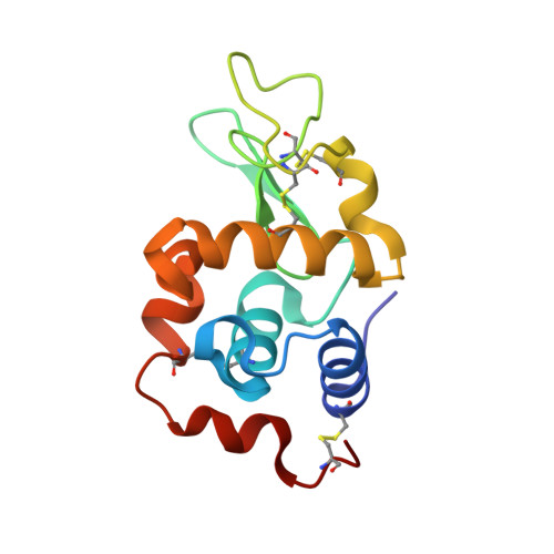
    The anticancer agents cisplatin and carboplatin bind to histidine in a protein. This crystal structure study at data-collection temperatures of 100 and 300 K examines their relative binding affinities to a histidine side chain and the effect of a high X-ray radiation dose of up to ∼1.8 MGy on the stability of the subsequent protein-Pt adducts. Cisplatin binding is visible at the histidine residue, but carboplatin binding is not. Five refined X-ray crystal structures are presented: one at 100 K as a reference and four at 300 K. The diffraction resolutions are 1.8, 2.0, 2.8, 2.9 and 3.5 Å.


  • Organizational Affiliation

    School of Chemistry, University of Manchester, England, UK. john.helliwell@manchester.ac.uk


Macromolecules
Find similar proteins by:  (by identity cutoff)  |  3D Structure
Entity ID: 1
MoleculeChains Sequence LengthOrganismDetailsImage
Lysozyme C129Gallus gallusMutation(s): 0 
EC: 3.2.1.17
UniProt
Find proteins for P00698 (Gallus gallus)
Explore P00698 
Go to UniProtKB:  P00698
Entity Groups  
Sequence Clusters30% Identity50% Identity70% Identity90% Identity95% Identity100% Identity
UniProt GroupP00698
Sequence Annotations
Expand
  • Reference Sequence
Experimental Data & Validation

Experimental Data

  • Method: X-RAY DIFFRACTION
  • Resolution: 3.50 Å
  • R-Value Free: 0.235 
  • R-Value Work: 0.170 
  • R-Value Observed: 0.172 
  • Space Group: P 43 21 2
Unit Cell:
Length ( Å )Angle ( ˚ )
a = 79.87α = 90
b = 79.87β = 90
c = 37.768γ = 90
Software Package:
Software NamePurpose
APEXdata collection
PHASERphasing
REFMACrefinement
APEXdata reduction

Structure Validation

View Full Validation Report


Entry History 

Deposition Data

Revision History  (Full details and data files)

  • Version 1.0: 2013-01-02
    Type: Initial release
  • Version 1.1: 2013-01-23
    Changes: Database references
  • Version 1.2: 2023-09-13
    Changes: Data collection, Database references, Derived calculations, Refinement description