4G8M | pdb_00004g8m

Crystal structure of the GluA2 ligand-binding domain (S1S2J) in complex with the agonist CBG-IV at 2.05A resolution


Experimental Data Snapshot

  • Method: X-RAY DIFFRACTION
  • Resolution: 2.05 Å
  • R-Value Free: 
    0.228 (Depositor), 0.208 (DCC) 
  • R-Value Work: 
    0.180 (Depositor) 
  • R-Value Observed: 
    0.182 (Depositor) 

Starting Model: experimental
View more details

wwPDB Validation 3D Report Full Report

Validation slider image for 4G8M

Ligand Structure Quality Assessment 


This is version 1.4 of the entry. See complete history

Literature

Pharmacological and structural characterization of conformationally restricted (S)-glutamate analogues at ionotropic glutamate receptors.

Juknaite, L.Venskutonyte, R.Assaf, Z.Faure, S.Gefflaut, T.Aitken, D.J.Nielsen, B.Gajhede, M.Kastrup, J.S.Bunch, L.Frydenvang, K.Pickering, D.S.

(2012) J Struct Biol 180: 39-46

  • DOI: https://doi.org/10.1016/j.jsb.2012.07.001
  • Primary Citation Related Structures: 
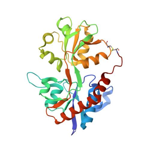
    4G8M, 4G8N

  • PubMed Abstract: 

    Conformationally restricted glutamate analogues have been pharmacologically characterized at AMPA and kainate receptors and the crystal structures have been solved of the ligand (2S,1'R,2'S)-2-(2'-carboxycyclobutyl)glycine (CBG-IV) in complex with the ligand binding domains of the AMPA receptor GluA2 and the kainate receptor GluK3. These structures show that CBG-IV interacts with the binding pocket in the same way as (S)-glutamate. The binding affinities reveal that CBG-IV has high affinity at the AMPA and kainate receptor subtypes. Appreciable binding affinity of CBG-IV was not observed at NMDA receptors, where the introduction of the carbocyclic ring is expected to lead to a steric clash with binding site residues. CBG-IV was demonstrated to be an agonist at both GluA2 and the kainate receptor GluK1. CBG-IV showed high affinity binding to GluK1 compared to GluA2, GluK2 and GluK3, which exhibited lower affinity for CBG-IV. The structure of GluA2 LBD and GluK3 LBD in complex with CBG-IV revealed similar binding site interactions to those of (S)-glutamate. No major conformational rearrangements compared to the (S)-glutamate bound conformation were found in GluK3 in order to accommodate CBG-IV, in contrast with GluA2 where a shift in lobe D2 binding site residues occurs, leading to an increased binding cavity volume compared to the (S)-glutamate bound structure.


  • Organizational Affiliation
    • Department of Drug Design and Pharmacology, Faculty of Health and Medical Sciences, University of Copenhagen, Universitetsparken 2, DK-2100 Copenhagen, Denmark.

Macromolecule Content 

  • Total Structure Weight: 59.75 kDa 
  • Atom Count: 4,550 
  • Modeled Residue Count: 523 
  • Deposited Residue Count: 526 
  • Unique protein chains: 1

Macromolecules

Find similar proteins by:|  3D Structure
Entity ID: 1
MoleculeChains  Sequence LengthOrganismDetailsImage
Glutamate receptor 2
A, B
263Rattus norvegicusMutation(s): 0 
Gene Names: Glur2Gria2
UniProt
Find proteins for P19491 (Rattus norvegicus)
Explore P19491 
Go to UniProtKB:  P19491
Entity Groups
Sequence Clusters30% Identity50% Identity70% Identity90% Identity95% Identity100% Identity
UniProt GroupP19491
Sequence Annotations
Expand
Reference Sequence

Small Molecules

Ligands 4 Unique
IDChains Name / Formula / InChI Key2D Diagram3D Interactions
G8M

Query on G8M


Download:Ideal Coordinates CCD File
C [auth A],
J [auth B]
(1S,2R)-2-[(S)-amino(carboxy)methyl]cyclobutanecarboxylic acid
C7 H11 N O4
SRAFHGOPGVYULO-WISUUJSJSA-N
SO4

Query on SO4


Download:Ideal Coordinates CCD File
D [auth A]
E [auth A]
F [auth A]
G [auth A]
K [auth B]
D [auth A],
E [auth A],
F [auth A],
G [auth A],
K [auth B],
L [auth B],
M [auth B]
SULFATE ION
O4 S
QAOWNCQODCNURD-UHFFFAOYSA-L
GOL

Query on GOL


Download:Ideal Coordinates CCD File
I [auth A],
P [auth B]
GLYCEROL
C3 H8 O3
PEDCQBHIVMGVHV-UHFFFAOYSA-N
CL

Query on CL


Download:Ideal Coordinates CCD File
H [auth A],
N [auth B],
O [auth B]
CHLORIDE ION
Cl
VEXZGXHMUGYJMC-UHFFFAOYSA-M

Experimental Data & Validation

Experimental Data

  • Method: X-RAY DIFFRACTION
  • Resolution: 2.05 Å
  • R-Value Free:  0.228 (Depositor), 0.208 (DCC) 
  • R-Value Work:  0.180 (Depositor) 
  • R-Value Observed: 0.182 (Depositor) 
Space Group: P 21 21 2
Unit Cell:
Length ( Å )Angle ( ˚ )
a = 98.367α = 90
b = 120.926β = 90
c = 47.929γ = 90
Software Package:
Software NamePurpose
PHASERphasing
PHENIXrefinement
XDSdata reduction
SCALAdata scaling

Structure Validation

View Full Validation Report


Ligand Structure Quality Assessment 


Entry History 

Deposition Data

Revision History  (Full details and data files)

  • Version 1.0: 2012-08-08
    Type: Initial release
  • Version 1.1: 2012-11-14
    Changes: Database references
  • Version 1.2: 2017-08-23
    Changes: Data collection, Refinement description, Source and taxonomy
  • Version 1.3: 2023-09-13
    Changes: Data collection, Database references, Derived calculations, Refinement description
  • Version 1.4: 2024-11-06
    Changes: Structure summary