4G3T

Mycobacterium smegmatis DprE1 - hexagonal crystal form


Experimental Data Snapshot

  • Method: X-RAY DIFFRACTION
  • Resolution: 2.35 Å
  • R-Value Free: 0.256 
  • R-Value Work: 0.227 
  • R-Value Observed: 0.229 

wwPDB Validation   3D Report Full Report


This is version 1.2 of the entry. See complete history


Literature

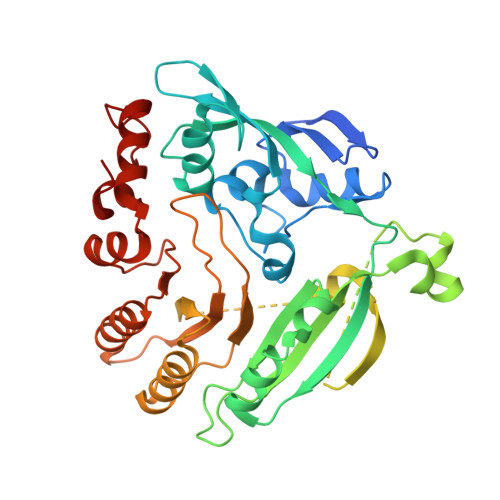
Crystal structure of decaprenylphosphoryl-beta- D-ribose 2'-epimerase from Mycobacterium smegmatis.

Li, H.Jogl, G.

(2013) Proteins 81: 538-543

  • DOI: https://doi.org/10.1002/prot.24220
  • Primary Citation of Related Structures:  
    4G3T, 4G3U

  • PubMed Abstract: 

    Decaprenylphosphoryl-β-D-ribose 2'-epimerase (DprE1) is an essential enzyme in the biosynthesis of cell wall components and a target for development of anti-tuberculosis drugs. We determined the crystal structure of a truncated form of DprE1 from Mycobacterium smegmatis in two crystal forms to up to 2.35 Å resolution. The structure extends from residue 75 to the C-terminus and shares homology with FAD-dependent oxidoreductases of the vanillyl-alcohol oxidase family including the DprE1 homologue from M. tuberculosis. The M. smegmatis DprE1 structure reported here provides further insights into the active site geometry of this tuberculosis drug target.


  • Organizational Affiliation

    Department of Molecular Biology, Cellular Biology and Biochemistry, Brown University, Providence, Rhode Island 02912, USA.


Macromolecules
Find similar proteins by:  (by identity cutoff)  |  3D Structure
Entity ID: 1
MoleculeChains Sequence LengthOrganismDetailsImage
oxidoreductase DprE1403Mycolicibacterium smegmatisMutation(s): 0 
Gene Names: MSMEG_6382
EC: 1
UniProt
Find proteins for A0R607 (Mycolicibacterium smegmatis (strain ATCC 700084 / mc(2)155))
Explore A0R607 
Go to UniProtKB:  A0R607
Entity Groups  
Sequence Clusters30% Identity50% Identity70% Identity90% Identity95% Identity100% Identity
UniProt GroupA0R607
Sequence Annotations
Expand
  • Reference Sequence
Experimental Data & Validation

Experimental Data

  • Method: X-RAY DIFFRACTION
  • Resolution: 2.35 Å
  • R-Value Free: 0.256 
  • R-Value Work: 0.227 
  • R-Value Observed: 0.229 
  • Space Group: P 32 2 1
Unit Cell:
Length ( Å )Angle ( ˚ )
a = 64.148α = 90
b = 64.148β = 90
c = 275.462γ = 120
Software Package:
Software NamePurpose
XSCALEdata scaling
PHENIXrefinement
PDB_EXTRACTdata extraction
CBASSdata collection
XDSdata reduction
XDSdata scaling
SnBphasing

Structure Validation

View Full Validation Report


Entry History 

Deposition Data

Revision History  (Full details and data files)

  • Version 1.0: 2012-12-05
    Type: Initial release
  • Version 1.1: 2013-08-28
    Changes: Database references
  • Version 1.2: 2024-02-28
    Changes: Data collection, Database references