4G1R | pdb_00004g1r

Crystal structure of anti-HIV actinohivin in complex with alphs-1,2-mannobiose (Form II)


Experimental Data Snapshot

  • Method: X-RAY DIFFRACTION
  • Resolution: 1.57 Å
  • R-Value Free: 
    0.194 (Depositor), 0.210 (DCC) 
  • R-Value Work: 
    0.160 (Depositor), 0.170 (DCC) 
  • R-Value Observed: 
    0.162 (Depositor) 

Starting Model: experimental
View more details

wwPDB Validation   3D Report Full Report


This is version 2.3 of the entry. See complete history


Literature

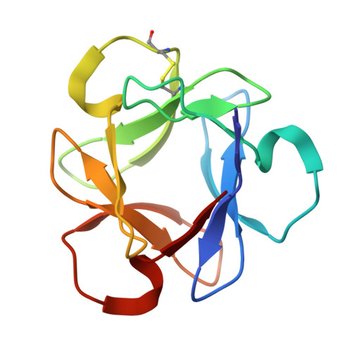
Peculiarity in crystal packing of anti-HIV lectin actinohivin in complex with alpha (1-2)mannobiose.

Suzuki, K.Tsunoda, M.Hoque, M.M.Zhang, F.Jiang, J.Zhang, X.Ohbayashi, N.Tanaka, H.Takenaka, A.

(2013) Acta Crystallogr D Biol Crystallogr 69: 1818-1825

  • DOI: https://doi.org/10.1107/S0907444913017812
  • Primary Citation of Related Structures:  
    4END, 4G1R

  • PubMed Abstract: 

    Previously, the anti-HIV lectin actinohivin (AH) was cocrystallized with the target α(1-2)mannobiose (MB) in the apparent space group P213. However, three MB-bound AH rotamers generated by ±120° rotations around the molecular pseudo-threefold rotation axis are packed randomly in the unit cell according to P212121 symmetry [Hoque et al. (2012). Acta Cryst. D68, 1671-1679]. It was found that the AH used for crystallization contains short peptides attached to the N-terminus [Suzuki et al. (2012). Acta Cryst. F68, 1060-1063], which cause packing disorder. In the present study, the fully mature homogeneous AH has been cocrystallized with MB into two new crystal forms at different pH. X-ray analyses of the two forms reveal that they have peculiar character in that the space groups are the same, P22121, and the unit-cell parameters are almost the same with the exception of the length of the a axis, which is doubled in one form. The use of homogeneous AH resulted in the absence of disorder in both crystals and an improvement in the resolution, thereby establishing the basis for AH binding to the target MB. In addition, the two crystal structures clarify the interaction modes between AH molecules, which is important knowledge for understanding the multiple binding effect generated when two AH molecules are linked together with a short peptide [Takahashi et al. (2011). J. Antibiot. 64, 551-557].


  • Organizational Affiliation
    • College of Science and Engineering, Iwaki Meisei University, Iwaki, Fukushima 970-8551, Japan.

Macromolecules
Find similar proteins by:  (by identity cutoff)  |  3D Structure
Entity ID: 1
MoleculeChains Sequence LengthOrganismDetailsImage
ActinohivinA,
B [auth C]
114actinomycete K97-0003Mutation(s): 0 
UniProt
Find proteins for Q9KWN0 (Actinomycete sp. (strain K97-0003))
Explore Q9KWN0 
Go to UniProtKB:  Q9KWN0
Entity Groups  
Sequence Clusters30% Identity50% Identity70% Identity90% Identity95% Identity100% Identity
UniProt GroupQ9KWN0
Sequence Annotations
Expand
  • Reference Sequence
Oligosaccharides

Help

Entity ID: 2
MoleculeChains Length2D Diagram Glycosylation3D Interactions
alpha-D-mannopyranose-(1-2)-alpha-D-mannopyranose
C [auth B],
D,
E,
F,
G,
C [auth B],
D,
E,
F,
G,
H
2N/A
Glycosylation Resources
GlyTouCan:  G53402KW
GlyCosmos:  G53402KW
GlyGen:  G53402KW
Experimental Data & Validation

Experimental Data

  • Method: X-RAY DIFFRACTION
  • Resolution: 1.57 Å
  • R-Value Free:  0.194 (Depositor), 0.210 (DCC) 
  • R-Value Work:  0.160 (Depositor), 0.170 (DCC) 
  • R-Value Observed: 0.162 (Depositor) 
Space Group: P 2 21 21
Unit Cell:
Length ( Å )Angle ( ˚ )
a = 54.76α = 90
b = 66.86β = 90
c = 66.86γ = 90
Software Package:
Software NamePurpose
HKL-2000data collection
AMoREphasing
REFMACrefinement
MOSFLMdata reduction
SCALAdata scaling

Structure Validation

View Full Validation Report


Entry History 

Deposition Data

Revision History  (Full details and data files)

  • Version 1.0: 2013-07-17
    Type: Initial release
  • Version 2.0: 2020-07-29
    Type: Remediation
    Reason: Carbohydrate remediation
    Changes: Atomic model, Data collection, Derived calculations, Non-polymer description, Structure summary
  • Version 2.1: 2023-11-08
    Changes: Data collection, Database references, Refinement description, Structure summary
  • Version 2.2: 2024-11-20
    Changes: Structure summary
  • Version 2.3: 2025-08-06
    Changes: Database references