4G09 | pdb_00004g09

The crystal structure of the C366S mutant of HDH from Brucella suis in complex with a substituted benzyl ketone


Experimental Data Snapshot

  • Method: X-RAY DIFFRACTION
  • Resolution: 1.90 Å
  • R-Value Free: 
    0.253 (Depositor), 0.253 (DCC) 
  • R-Value Work: 
    0.213 (Depositor), 0.213 (DCC) 
  • R-Value Observed: 
    0.213 (Depositor) 

wwPDB Validation 3D Report Full Report

Validation slider image for 4G09

Ligand Structure Quality Assessment 


This is version 1.3 of the entry. See complete history

Literature

Structural basis for the rational design of new anti-Brucella agents: The crystal structure of the C366S mutant of l-histidinol dehydrogenase from Brucella suis.

D'ambrosio, K.Lopez, M.Dathan, N.A.Ouahrani-Bettache, S.Kohler, S.Ascione, G.Monti, S.M.Winum, J.Y.De Simone, G.

(2014) Biochimie 97: 114-120

  • DOI: https://doi.org/10.1016/j.biochi.2013.09.028
  • Primary Citation Related Structures: 
    4G07, 4G09

  • PubMed Abstract: 

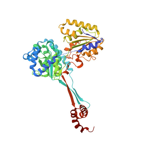
    L-Histidinol dehydrogenase from Brucella suis (BsHDH) is an enzyme involved in the histidine biosynthesis pathway which is absent in mammals, thus representing a very interesting target for the development of anti-Brucella agents. In this paper we report the crystallographic structure of a mutated form of BsHDH both in its unbound form and in complex with a nanomolar inhibitor. These studies provide the first structural background for the rational design of potent HDH inhibitors, thus offering new hints for clinical applications.


  • Organizational Affiliation
    • Istituto di Biostrutture e Bioimmagini-CNR, Via Mezzocannone 16, 80134 Napoli, Italy.

Macromolecule Content 

  • Total Structure Weight: 47.82 kDa 
  • Atom Count: 3,544 
  • Modeled Residue Count: 432 
  • Deposited Residue Count: 438 
  • Unique protein chains: 1

Macromolecules

Find similar proteins by:|  3D Structure
Entity ID: 1
MoleculeChains  Sequence LengthOrganismDetailsImage
Histidinol dehydrogenase438Brucella suis 1330Mutation(s): 1 
Gene Names: hisDBR0252BS1330_I0253
EC: 1.1.1.23
UniProt
Find proteins for Q8G2R2 (Brucella suis biovar 1 (strain 1330))
Explore Q8G2R2 
Go to UniProtKB:  Q8G2R2
Entity Groups
Sequence Clusters30% Identity50% Identity70% Identity90% Identity95% Identity100% Identity
UniProt GroupQ8G2R2
Sequence Annotations
Expand
Reference Sequence

Small Molecules

Experimental Data & Validation

Experimental Data

  • Method: X-RAY DIFFRACTION
  • Resolution: 1.90 Å
  • R-Value Free:  0.253 (Depositor), 0.253 (DCC) 
  • R-Value Work:  0.213 (Depositor), 0.213 (DCC) 
  • R-Value Observed: 0.213 (Depositor) 
Space Group: C 2 2 21
Unit Cell:
Length ( Å )Angle ( ˚ )
a = 76.654α = 90
b = 105.072β = 90
c = 111.25γ = 90
Software Package:
Software NamePurpose
CNSrefinement
DENZOdata reduction
HKL-2000data scaling
CNSphasing

Structure Validation

View Full Validation Report


Ligand Structure Quality Assessment 


Entry History 

Deposition Data

Revision History  (Full details and data files)

  • Version 1.0: 2013-10-02
    Type: Initial release
  • Version 1.1: 2013-11-06
    Changes: Database references
  • Version 1.2: 2014-02-05
    Changes: Database references
  • Version 1.3: 2024-02-28
    Changes: Data collection, Database references, Derived calculations