4FSL | pdb_00004fsl

Crystal structure of beta-site app-cleaving enzyme 1 (BACE-DB-MUT) complex with N-(N-(4- acetamido-3-chloro-5-methylbenzyl)carbamimidoyl)-3-(4- methoxyphenyl)-5-methyl-4-isothiazolecarboxamide


Experimental Data Snapshot

  • Method: X-RAY DIFFRACTION
  • Resolution: 2.50 Å
  • R-Value Free: 
    0.277 (Depositor), 0.274 (DCC) 
  • R-Value Work: 
    0.224 (Depositor), 0.223 (DCC) 
  • R-Value Observed: 
    0.227 (Depositor) 

wwPDB Validation 3D Report Full Report

Validation slider image for 4FSL

Ligand Structure Quality Assessment 


This is version 1.5 of the entry. See complete history

Literature

Acyl Guanidine Inhibitors of beta-Secretase (BACE-1): Optimization of a Micromolar Hit to a Nanomolar Lead via Iterative Solid- and Solution-Phase Library Synthesis

Gerritz, S.W.Zhai, W.Shi, S.Zhu, S.Toyn, J.H.Meredith, J.E.Iben, L.G.Burton, C.R.Albright, C.F.Good, A.C.Tebben, A.J.Muckelbauer, J.K.Camac, D.M.Metzler, W.Cook, L.S.Padmanabha, R.Lentz, K.A.Sofia, M.J.Poss, M.A.Macor, J.E.Thompson, L.A.

(2012) J Med Chem 55: 9208-9223

  • DOI: https://doi.org/10.1021/jm300931y
  • Primary Citation Related Structures: 
    4FSE, 4FSL

  • PubMed Abstract: 

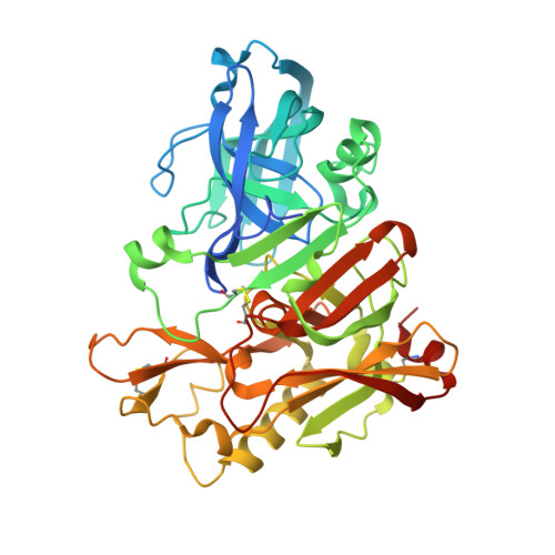
    This report describes the discovery and optimization of a BACE-1 inhibitor series containing an unusual acyl guanidine chemotype that was originally synthesized as part of a 6041-membered solid-phase library. The synthesis of multiple follow-up solid- and solution-phase libraries facilitated the optimization of the original micromolar hit into a single-digit nanomolar BACE-1 inhibitor in both radioligand binding and cell-based functional assay formats. The X-ray structure of representative inhibitors bound to BACE-1 revealed a number of key ligand:protein interactions, including a hydrogen bond between the side chain amide of flap residue Gln73 and the acyl guanidine carbonyl group, and a cation-π interaction between Arg235 and the isothiazole 4-methoxyphenyl substituent. Following subcutaneous administration in rats, an acyl guanidine inhibitor with single-digit nanomolar activity in cells afforded good plasma exposures and a dose-dependent reduction in plasma Aβ levels, but poor brain exposure was observed (likely due to Pgp-mediated efflux), and significant reductions in brain Aβ levels were not obtained.


  • Organizational Affiliation
    • Bristol-Myers Squibb Research, 5 Research Parkway, Wallingford, Connecticut 06492, United States. samuel.gerritz@bms.com

Macromolecule Content 

  • Total Structure Weight: 187.16 kDa 
  • Atom Count: 13,119 
  • Modeled Residue Count: 1,548 
  • Deposited Residue Count: 1,648 
  • Unique protein chains: 1

Macromolecules

Find similar proteins by:|  3D Structure
Entity ID: 1
MoleculeChains  Sequence LengthOrganismDetailsImage
Beta-secretase 1A,
B,
C [auth D],
D [auth E]
412Homo sapiensMutation(s): 0 
Gene Names: BACE1BACEKIAA1149
EC: 3.4.23.46
UniProt & NIH Common Fund Data Resources
Find proteins for P56817 (Homo sapiens)
Explore P56817 
Go to UniProtKB:  P56817
PHAROS:  P56817
GTEx:  ENSG00000186318 
Entity Groups
Sequence Clusters30% Identity50% Identity70% Identity90% Identity95% Identity100% Identity
UniProt GroupP56817
Sequence Annotations
Expand
Reference Sequence

Small Molecules

Ligands 2 Unique
IDChains Name / Formula / InChI Key2D Diagram3D Interactions
0VB

Query on 0VB


Download:Ideal Coordinates CCD File
E [auth A],
I [auth B],
M [auth D],
Q [auth E]
N-{N-[4-(acetylamino)-3-chloro-5-methylbenzyl]carbamimidoyl}-3-(4-methoxyphenyl)-5-methyl-1,2-thiazole-4-carboxamide
C23 H24 Cl N5 O3 S
MQPJKTGBSCCWTI-UHFFFAOYSA-N
IOD

Query on IOD


Download:Ideal Coordinates CCD File
F [auth A]
G [auth A]
H [auth A]
J [auth B]
K [auth B]
F [auth A],
G [auth A],
H [auth A],
J [auth B],
K [auth B],
L [auth B],
N [auth D],
O [auth D],
P [auth D],
R [auth E],
S [auth E],
T [auth E]
IODIDE ION
I
XMBWDFGMSWQBCA-UHFFFAOYSA-M

Experimental Data & Validation

Experimental Data

  • Method: X-RAY DIFFRACTION
  • Resolution: 2.50 Å
  • R-Value Free:  0.277 (Depositor), 0.274 (DCC) 
  • R-Value Work:  0.224 (Depositor), 0.223 (DCC) 
  • R-Value Observed: 0.227 (Depositor) 
Space Group: P 1 21 1
Unit Cell:
Length ( Å )Angle ( ˚ )
a = 86.54α = 90
b = 131.177β = 97.61
c = 90.315γ = 90
Software Package:
Software NamePurpose
REFMACrefinement
SCALEPACKdata scaling
CNSrefinement
DENZOdata reduction
PDB_EXTRACTdata extraction
HKL-2000data reduction
HKL-2000data scaling
CNSphasing

Structure Validation

View Full Validation Report


Ligand Structure Quality Assessment 


Entry History 

Deposition Data

Revision History  (Full details and data files)

  • Version 1.0: 2012-10-10
    Type: Initial release
  • Version 1.1: 2013-07-10
    Changes: Database references
  • Version 1.2: 2017-11-15
    Changes: Refinement description
  • Version 1.3: 2018-06-13
    Changes: Data collection
  • Version 1.4: 2019-07-17
    Changes: Data collection, Refinement description
  • Version 1.5: 2024-10-16
    Changes: Data collection, Database references, Derived calculations, Structure summary