4FMD | pdb_00004fmd

EspG-Rab1 complex structure at 3.05 A


Experimental Data Snapshot

  • Method: X-RAY DIFFRACTION
  • Resolution: 3.05 Å
  • R-Value Free: 
    0.266 (Depositor), 0.254 (DCC) 
  • R-Value Work: 
    0.212 (Depositor), 0.205 (DCC) 
  • R-Value Observed: 
    0.214 (Depositor) 

wwPDB Validation 3D Report Full Report

Validation slider image for 4FMD

Ligand Structure Quality Assessment 


This is version 1.2 of the entry. See complete history

Literature

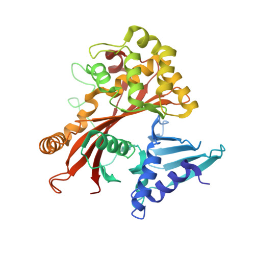
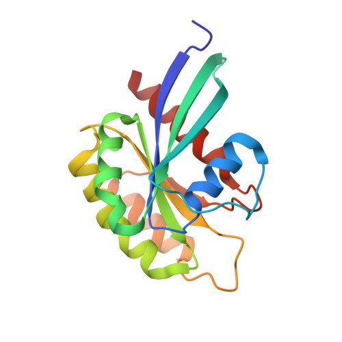
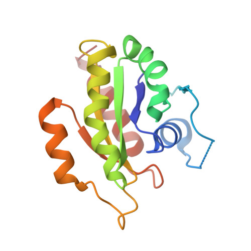
Structurally Distinct Bacterial TBC-like GAPs Link Arf GTPase to Rab1 Inactivation to Counteract Host Defenses.

Dong, N.Zhu, Y.Lu, Q.Hu, L.Zheng, Y.Shao, F.

(2012) Cell 150: 1029-1041

  • DOI: https://doi.org/10.1016/j.cell.2012.06.050
  • Primary Citation Related Structures: 
    4FMA, 4FMB, 4FMC, 4FMD, 4FME

  • PubMed Abstract: 

    Rab GTPases are frequent targets of vacuole-living bacterial pathogens for appropriate trafficking of the vacuole. Here we discover that bacterial effectors including VirA from nonvacuole Shigella flexneri and EspG from extracellular Enteropathogenic Escherichia coli (EPEC) harbor TBC-like dual-finger motifs and exhibits potent RabGAP activities. Specific inactivation of Rab1 by VirA/EspG disrupts ER-to-Golgi trafficking. S. flexneri intracellular persistence requires VirA TBC-like GAP activity that mediates bacterial escape from autophagy-mediated host defense. Rab1 inactivation by EspG severely blocks host secretory pathway, resulting in inhibited interleukin-8 secretion from infected cells. Crystal structures of VirA/EspG-Rab1-GDP-aluminum fluoride complexes highlight TBC-like catalytic role for the arginine and glutamine finger residues and reveal a 3D architecture distinct from that of the TBC domain. Structure of Arf6-EspG-Rab1 ternary complex illustrates a pathogenic signaling complex that rewires host Arf signaling to Rab1 inactivation. Structural distinctions of VirA/EspG further predict a possible extensive presence of TBC-like RabGAP effectors in counteracting various host defenses.


  • Organizational Affiliation
    • National Institute of Biological Sciences, Beijing 102206, China.

Macromolecule Content 

  • Total Structure Weight: 176.12 kDa 
  • Atom Count: 12,146 
  • Modeled Residue Count: 1,543 
  • Deposited Residue Count: 1,559 
  • Unique protein chains: 3

Macromolecules

Find similar proteins by:|  3D Structure
Entity ID: 1
MoleculeChains  Sequence LengthOrganismDetailsImage
EspG protein
A, C, E
351Escherichia coliMutation(s): 0 
Gene Names: espG
UniProt
Find proteins for Q7DB50 (Escherichia coli O157:H7)
Explore Q7DB50 
Go to UniProtKB:  Q7DB50
Entity Groups
Sequence Clusters30% Identity50% Identity70% Identity90% Identity95% Identity100% Identity
UniProt GroupQ7DB50
Sequence Annotations
Expand
Reference Sequence
Find similar proteins by:|  3D Structure
Entity ID: 2
MoleculeChains  Sequence LengthOrganismDetailsImage
Ras-related protein Rab-1A
B, D
171Homo sapiensMutation(s): 0 
Gene Names: RAB1RAB1A
EC: 3.6.5.2
UniProt & NIH Common Fund Data Resources
Find proteins for P62820 (Homo sapiens)
Explore P62820 
Go to UniProtKB:  P62820
PHAROS:  P62820
GTEx:  ENSG00000138069 
Entity Groups
Sequence Clusters30% Identity50% Identity70% Identity90% Identity95% Identity100% Identity
UniProt GroupP62820
Sequence Annotations
Expand
Reference Sequence
Find similar proteins by:|  3D Structure
Entity ID: 3
MoleculeChains  Sequence LengthOrganismDetailsImage
Ras-related protein Rab-1A164Homo sapiensMutation(s): 0 
Gene Names: RAB1RAB1A
EC: 3.6.5.2
UniProt & NIH Common Fund Data Resources
Find proteins for P62820 (Homo sapiens)
Explore P62820 
Go to UniProtKB:  P62820
PHAROS:  P62820
GTEx:  ENSG00000138069 
Entity Groups
Sequence Clusters30% Identity50% Identity70% Identity90% Identity95% Identity100% Identity
UniProt GroupP62820
Sequence Annotations
Expand
Reference Sequence

Small Molecules

Ligands 5 Unique
IDChains Name / Formula / InChI Key2D Diagram3D Interactions
GDP

Query on GDP


Download:Ideal Coordinates CCD File
H [auth B],
L [auth D],
P [auth F]
GUANOSINE-5'-DIPHOSPHATE
C10 H15 N5 O11 P2
QGWNDRXFNXRZMB-UUOKFMHZSA-N
PGE

Query on PGE


Download:Ideal Coordinates CCD File
J [auth C]TRIETHYLENE GLYCOL
C6 H14 O4
ZIBGPFATKBEMQZ-UHFFFAOYSA-N
PEG

Query on PEG


Download:Ideal Coordinates CCD File
N [auth E]DI(HYDROXYETHYL)ETHER
C4 H10 O3
MTHSVFCYNBDYFN-UHFFFAOYSA-N
AF3

Query on AF3


Download:Ideal Coordinates CCD File
G [auth B],
K [auth D],
O [auth F]
ALUMINUM FLUORIDE
Al F3
KLZUFWVZNOTSEM-UHFFFAOYSA-K
MG

Query on MG


Download:Ideal Coordinates CCD File
I [auth B],
M [auth D],
Q [auth F]
MAGNESIUM ION
Mg
JLVVSXFLKOJNIY-UHFFFAOYSA-N

Experimental Data & Validation

Experimental Data

  • Method: X-RAY DIFFRACTION
  • Resolution: 3.05 Å
  • R-Value Free:  0.266 (Depositor), 0.254 (DCC) 
  • R-Value Work:  0.212 (Depositor), 0.205 (DCC) 
  • R-Value Observed: 0.214 (Depositor) 
Space Group: I 2 2 2
Unit Cell:
Length ( Å )Angle ( ˚ )
a = 106.568α = 90
b = 153.221β = 90
c = 230.805γ = 90
Software Package:
Software NamePurpose
SCALEPACKdata scaling
REFMACrefinement
PDB_EXTRACTdata extraction
DENZOdata reduction
PHASESphasing

Structure Validation

View Full Validation Report


Ligand Structure Quality Assessment 


Entry History 

Deposition Data

Revision History  (Full details and data files)

  • Version 1.0: 2012-09-05
    Type: Initial release
  • Version 1.1: 2012-10-03
    Changes: Database references
  • Version 1.2: 2024-02-28
    Changes: Data collection, Database references, Derived calculations