4FKQ | pdb_00004fkq

Crystal structure of the cdk2 in complex with oxindole inhibitor


Experimental Data Snapshot

  • Method: X-RAY DIFFRACTION
  • Resolution: 1.75 Å
  • R-Value Free: 
    0.195 (Depositor), 0.199 (DCC) 
  • R-Value Work: 
    0.176 (Depositor), 0.174 (DCC) 
  • R-Value Observed: 
    0.177 (Depositor) 

Starting Model: experimental
View more details

wwPDB Validation 3D Report Full Report

Validation slider image for 4FKQ

Ligand Structure Quality Assessment 


This is version 1.4 of the entry. See complete history

Literature

Crystal structure of the cdk2 in complex with oxindole inhibitor

Kang, Y.N.Stuckey, J.A.

To be published.

Macromolecule Content 

  • Total Structure Weight: 34.45 kDa 
  • Atom Count: 2,570 
  • Modeled Residue Count: 284 
  • Deposited Residue Count: 299 
  • Unique protein chains: 1

Macromolecules

Find similar proteins by:|  3D Structure
Entity ID: 1
MoleculeChains  Sequence LengthOrganismDetailsImage
Cyclin-dependent kinase 2299Homo sapiensMutation(s): 1 
Gene Names: CDK2CDKN2
EC: 2.7.11.22
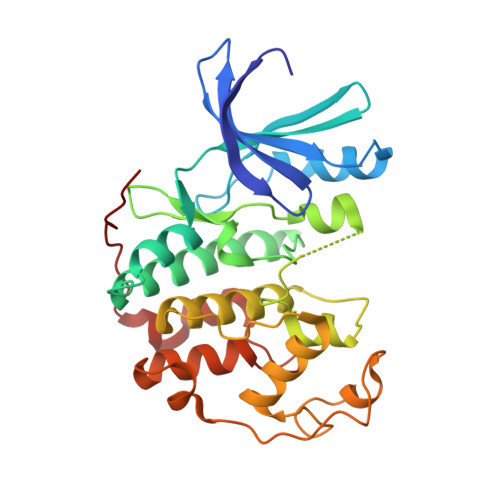
UniProt & NIH Common Fund Data Resources
Find proteins for P24941 (Homo sapiens)
Explore P24941 
Go to UniProtKB:  P24941
PHAROS:  P24941
GTEx:  ENSG00000123374 
Entity Groups
Sequence Clusters30% Identity50% Identity70% Identity90% Identity95% Identity100% Identity
UniProt GroupP24941
Sequence Annotations
Expand
Reference Sequence

Small Molecules

Ligands 2 Unique
IDChains Name / Formula / InChI Key2D Diagram3D Interactions
42K

Query on 42K


Download:Ideal Coordinates CCD File
B [auth A]4-[(2Z)-2-(7-oxidanylidene-3,6-dihydropyrrolo[3,2-e]benzotriazol-8-ylidene)hydrazinyl]benzenesulfonamide
C14 H11 N7 O3 S
AQAPFJBZSZVEGI-UHFFFAOYSA-N
GOL

Query on GOL


Download:Ideal Coordinates CCD File
C [auth A]GLYCEROL
C3 H8 O3
PEDCQBHIVMGVHV-UHFFFAOYSA-N
Binding Affinity Annotations 
IDSourceBinding Affinity
42K BindingDB:  4FKQ Kd: min: 191, max: 528 (nM) from 2 assay(s)
ΔG: min: -3.82e+1, max: -7.78e+0 (kJ/mol) from 6 assay(s)

Experimental Data & Validation

Experimental Data

  • Method: X-RAY DIFFRACTION
  • Resolution: 1.75 Å
  • R-Value Free:  0.195 (Depositor), 0.199 (DCC) 
  • R-Value Work:  0.176 (Depositor), 0.174 (DCC) 
  • R-Value Observed: 0.177 (Depositor) 
Space Group: P 21 21 21
Unit Cell:
Length ( Å )Angle ( ˚ )
a = 53.361α = 90
b = 72.112β = 90
c = 72.402γ = 90
Software Package:
Software NamePurpose
MD2data collection
PHASERphasing
BUSTERrefinement
HKL-2000data reduction
HKL-2000data scaling

Structure Validation

View Full Validation Report


Ligand Structure Quality Assessment 


Entry History 

Deposition Data

Revision History  (Full details and data files)

  • Version 1.0: 2013-05-08
    Type: Initial release
  • Version 1.1: 2013-05-22
    Changes: Other
  • Version 1.2: 2016-11-30
    Changes: Non-polymer description
  • Version 1.3: 2023-09-13
    Changes: Data collection, Database references, Derived calculations, Refinement description
  • Version 1.4: 2024-11-27
    Changes: Structure summary