4FCM

Crystal structure of the NTF2-like domain of human G3BP1 in complex with a peptide


Experimental Data Snapshot

  • Method: X-RAY DIFFRACTION
  • Resolution: 2.69 Å
  • R-Value Free: 0.282 
  • R-Value Work: 0.225 
  • R-Value Observed: 0.228 

wwPDB Validation   3D Report Full Report


This is version 1.4 of the entry. See complete history


Literature

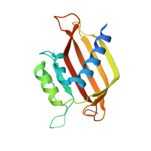
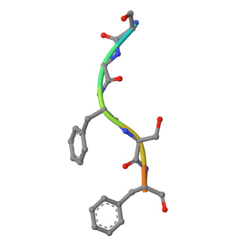
Crystal Structures of the Human G3BP1 NTF2-Like Domain Visualize FxFG Nup Repeat Specificity.

Vognsen, T.Moller, I.R.Kristensen, O.

(2013) PLoS One 8: e80947-e80947

  • DOI: https://doi.org/10.1371/journal.pone.0080947
  • Primary Citation of Related Structures:  
    4FCJ, 4FCM, 4IIA

  • PubMed Abstract: 

    Ras GTPase Activating Protein SH3 Domain Binding Protein (G3BP) is a potential anti-cancer drug target implicated in several cellular functions. We have used protein crystallography to solve crystal structures of the human G3BP1 NTF2-like domain both alone and in complex with an FxFG Nup repeat peptide. Despite high structural similarity, the FxFG binding site is located between two alpha helices in the G3BP1 NTF2-like domain and not at the dimer interface as observed for nuclear transport factor 2. ITC studies showed specificity towards the FxFG motif but not FG and GLFG motifs. The unliganded form of the G3BP1 NTF2-like domain was solved in two crystal forms to resolutions of 1.6 and 3.3 Å in space groups P212121 and P6322 based on two different constructs, residues 1-139 and 11-139, respectively. Crystal packing of the N-terminal residues against a symmetry related molecule in the P212121 crystal form might indicate a novel ligand binding site that, however, remains to be validated. The crystal structures give insight into the nuclear transportation mechanisms of G3BP and provide a basis for future structure based drug design.


  • Organizational Affiliation

    Biostructural Research, Department of Drug Design and Pharmacology, Faculty of Health Sciences, University of Copenhagen, Copenhagen, Denmark.


Macromolecules
Find similar proteins by:  (by identity cutoff)  |  3D Structure
Entity ID: 1
MoleculeChains Sequence LengthOrganismDetailsImage
Ras GTPase-activating protein-binding protein 1
A, B
142Homo sapiensMutation(s): 0 
Gene Names: G3BP1G3BP
EC: 3.6.4.12 (PDB Primary Data), 3.6.4.13 (PDB Primary Data)
UniProt & NIH Common Fund Data Resources
Find proteins for Q13283 (Homo sapiens)
Explore Q13283 
Go to UniProtKB:  Q13283
PHAROS:  Q13283
GTEx:  ENSG00000145907 
Entity Groups  
Sequence Clusters30% Identity50% Identity70% Identity90% Identity95% Identity100% Identity
UniProt GroupQ13283
Sequence Annotations
Expand
  • Reference Sequence

Find similar proteins by:  Sequence   |   3D Structure  

Entity ID: 2
MoleculeChains Sequence LengthOrganismDetailsImage
Nucleoporin repeat peptide
C, D
9N/AMutation(s): 0 
Sequence Annotations
Expand
  • Reference Sequence
Small Molecules
Ligands 1 Unique
IDChains Name / Formula / InChI Key2D Diagram3D Interactions
PO4
Query on PO4

Download Ideal Coordinates CCD File 
E [auth A]PHOSPHATE ION
O4 P
NBIIXXVUZAFLBC-UHFFFAOYSA-K
Experimental Data & Validation

Experimental Data

  • Method: X-RAY DIFFRACTION
  • Resolution: 2.69 Å
  • R-Value Free: 0.282 
  • R-Value Work: 0.225 
  • R-Value Observed: 0.228 
  • Space Group: P 21 21 21
Unit Cell:
Length ( Å )Angle ( ˚ )
a = 42.78α = 90
b = 71.49β = 90
c = 89.5γ = 90
Software Package:
Software NamePurpose
MAR345data collection
PHENIXmodel building
PHENIXrefinement
XDSdata reduction
XSCALEdata scaling
PHENIXphasing

Structure Validation

View Full Validation Report


Entry History 

Deposition Data

Revision History  (Full details and data files)

  • Version 1.0: 2013-06-05
    Type: Initial release
  • Version 1.1: 2013-12-18
    Changes: Database references
  • Version 1.2: 2017-08-09
    Changes: Data collection, Database references
  • Version 1.3: 2017-11-15
    Changes: Refinement description
  • Version 1.4: 2023-09-13
    Changes: Data collection, Database references, Derived calculations, Refinement description