4F2J | pdb_00004f2j

Crystal structure of ZNF217 bound to DNA, P6522 crystal form


Experimental Data Snapshot

  • Method: X-RAY DIFFRACTION
  • Resolution: 2.64 Å
  • R-Value Free: 
    0.264 (Depositor), 0.254 (DCC) 
  • R-Value Work: 
    0.252 (Depositor), 0.249 (DCC) 
  • R-Value Observed: 
    0.253 (Depositor) 

wwPDB Validation 3D Report Full Report

Validation slider image for 4F2J

This is version 1.2 of the entry. See complete history

Literature

New insights into DNA recognition by zinc fingers revealed by structural analysis of the oncoprotein ZNF217

Vandevenne, M.S.Jacques, D.A.Artuz, C.Nguyen, C.D.Kwan, A.H.Segal, D.J.Matthews, J.M.Crossley, M.Guss, J.M.Mackay, J.P.

(2013) J Biological Chem 288: 10616-10627

  • DOI: https://doi.org/10.1074/jbc.M112.441451
  • Primary Citation Related Structures: 
    4F2J, 4IS1

  • PubMed Abstract: 

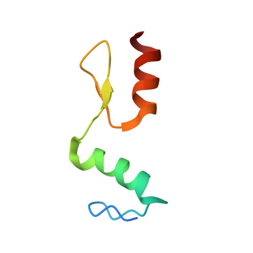
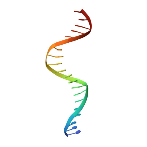
    Classical zinc fingers (ZFs) are one of the most abundant and best characterized DNA-binding domains. Typically, tandem arrays of three or more ZFs bind DNA target sequences with high affinity and specificity, and the mode of DNA recognition is sufficiently well understood that tailor-made ZF-based DNA-binding proteins can be engineered. We have shown previously that a two-zinc finger unit found in the transcriptional coregulator ZNF217 recognizes DNA but with an affinity and specificity that is lower than other ZF arrays. To investigate the basis for these differences, we determined the structure of a ZNF217-DNA complex. We show that although the overall position of the ZFs on the DNA closely resembles that observed for other ZFs, the side-chain interaction pattern differs substantially from the canonical model. The structure also reveals the presence of two methyl-π interactions, each featuring a tyrosine contacting a thymine methyl group. To our knowledge, interactions of this type have not previously been described in classical ZF-DNA complexes. Finally, we investigated the sequence specificity of this two-ZF unit and discuss how ZNF217 might discriminate its target DNA sites in the cell.


  • Organizational Affiliation
    • School of Molecular Bioscience, University of Sydney, New South Wales, 2006 Australia.

Macromolecule Content 

  • Total Structure Weight: 13.1 kDa 
  • Atom Count: 857 
  • Modeled Residue Count: 72 
  • Deposited Residue Count: 77 
  • Unique protein chains: 1
  • Unique nucleic acid chains: 1

Macromolecules


Find similar proteins by:|  3D Structure
Entity ID: 2
MoleculeChains  Sequence LengthOrganismDetailsImage
Zinc finger protein 217B [auth C]57Homo sapiensMutation(s): 0 
Gene Names: ZNF217ZABC1
UniProt & NIH Common Fund Data Resources
Find proteins for O75362 (Homo sapiens)
Explore O75362 
Go to UniProtKB:  O75362
PHAROS:  O75362
GTEx:  ENSG00000171940 
Entity Groups
Sequence Clusters30% Identity50% Identity70% Identity90% Identity95% Identity100% Identity
UniProt GroupO75362
Sequence Annotations
Expand
Reference Sequence
Find similar nucleic acids by:  Sequence
Entity ID: 1
MoleculeChains LengthOrganismImage
5'-D(*TP*TP*TP*GP*CP*AP*GP*AP*AP*TP*CP*GP*AP*TP*TP*CP*TP*GP*CP*A)-3'20N/A
Sequence Annotations
Expand
Reference Sequence

Experimental Data & Validation

Experimental Data

  • Method: X-RAY DIFFRACTION
  • Resolution: 2.64 Å
  • R-Value Free:  0.264 (Depositor), 0.254 (DCC) 
  • R-Value Work:  0.252 (Depositor), 0.249 (DCC) 
  • R-Value Observed: 0.253 (Depositor) 
Space Group: P 65 2 2
Unit Cell:
Length ( Å )Angle ( ˚ )
a = 56.928α = 90
b = 56.928β = 90
c = 242.542γ = 120
Software Package:
Software NamePurpose
DENZOdata reduction
SCALEPACKdata scaling
SOLVEphasing
RESOLVEphasing
REFMACrefinement
PDB_EXTRACTdata extraction
MAR345dtbdata collection

Structure Validation

View Full Validation Report


Entry History 

Deposition Data

Revision History  (Full details and data files)

  • Version 1.0: 2013-02-27
    Type: Initial release
  • Version 1.1: 2013-09-04
    Changes: Database references
  • Version 1.2: 2024-03-20
    Changes: Data collection, Database references, Derived calculations