4ERE | pdb_00004ere

crystal structure of MDM2 (17-111) in complex with compound 23


Experimental Data Snapshot

  • Method: X-RAY DIFFRACTION
  • Resolution: 1.80 Å
  • R-Value Free: 
    0.270 (Depositor), 0.290 (DCC) 
  • R-Value Work: 
    0.250 (Depositor), 0.277 (DCC) 

wwPDB Validation 3D Report Full Report

Validation slider image for 4ERE

Ligand Structure Quality Assessment 


This is version 1.2 of the entry. See complete history

Literature

Structure-based design of novel inhibitors of the MDM2-p53 interaction.

Rew, Y.Sun, D.Gonzalez-Lopez De Turiso, F.Bartberger, M.D.Beck, H.P.Canon, J.Chen, A.Chow, D.Deignan, J.Fox, B.M.Gustin, D.Huang, X.Jiang, M.Jiao, X.Jin, L.Kayser, F.Kopecky, D.J.Li, Y.Lo, M.C.Long, A.M.Michelsen, K.Oliner, J.D.Osgood, T.Ragains, M.Saiki, A.Y.Schneider, S.Toteva, M.Yakowec, P.Yan, X.Ye, Q.Yu, D.Zhao, X.Zhou, J.Medina, J.C.Olson, S.H.

(2012) J Med Chem 55: 4936-4954

  • DOI: https://doi.org/10.1021/jm300354j
  • Primary Citation Related Structures: 
    4ERE, 4ERF

  • PubMed Abstract: 

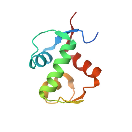
    Structure-based rational design led to the discovery of novel inhibitors of the MDM2-p53 protein-protein interaction. The affinity of these compounds for MDM2 was improved through conformational control of both the piperidinone ring and the appended N-alkyl substituent. Optimization afforded 29 (AM-8553), a potent and selective MDM2 inhibitor with excellent pharmacokinetic properties and in vivo efficacy.


  • Organizational Affiliation
    • Department of Therapeutic Discovery, Amgen Inc., 1120 Veterans Boulevard, South San Francisco, California 94080, United States.

Macromolecule Content 

  • Total Structure Weight: 23.45 kDa 
  • Atom Count: 1,667 
  • Modeled Residue Count: 175 
  • Deposited Residue Count: 192 
  • Unique protein chains: 1

Macromolecules

Find similar proteins by:|  3D Structure
Entity ID: 1
MoleculeChains  Sequence LengthOrganismDetailsImage
E3 ubiquitin-protein ligase Mdm2
A, B
96Homo sapiensMutation(s): 0 
Gene Names: MDM2
EC: 6.3.2 (PDB Primary Data), 2.3.2.27 (UniProt)
UniProt & NIH Common Fund Data Resources
Find proteins for Q00987 (Homo sapiens)
Explore Q00987 
Go to UniProtKB:  Q00987
PHAROS:  Q00987
GTEx:  ENSG00000135679 
Entity Groups
Sequence Clusters30% Identity50% Identity70% Identity90% Identity95% Identity100% Identity
UniProt GroupQ00987
Sequence Annotations
Expand
Reference Sequence

Small Molecules

Ligands 2 Unique
IDChains Name / Formula / InChI Key2D Diagram3D Interactions
0R2

Query on 0R2


Download:Ideal Coordinates CCD File
C [auth A],
E [auth B]
[(3R,5R,6S)-1-[(2S)-1-tert-butoxy-1-oxobutan-2-yl]-5-(3-chlorophenyl)-6-(4-chlorophenyl)-2-oxopiperidin-3-yl]acetic acid
C27 H31 Cl2 N O5
ZEMLYWMOSZRCQL-UWSZBUKDSA-N
SO4

Query on SO4


Download:Ideal Coordinates CCD File
D [auth A]SULFATE ION
O4 S
QAOWNCQODCNURD-UHFFFAOYSA-L
Binding Affinity Annotations 
IDSourceBinding Affinity
0R2 BindingDB:  4ERE IC50: min: 4, max: 40 (nM) from 2 assay(s)

Experimental Data & Validation

Experimental Data

  • Method: X-RAY DIFFRACTION
  • Resolution: 1.80 Å
  • R-Value Free:  0.270 (Depositor), 0.290 (DCC) 
  • R-Value Work:  0.250 (Depositor), 0.277 (DCC) 
Space Group: P 21 21 21
Unit Cell:
Length ( Å )Angle ( ˚ )
a = 43.15α = 90
b = 60.24β = 90
c = 68.09γ = 90
Software Package:
Software NamePurpose
CrystalCleardata collection
AMoREphasing
CNSrefinement
DENZOdata reduction
SCALEPACKdata scaling

Structure Validation

View Full Validation Report


Ligand Structure Quality Assessment 


Entry History 

Deposition Data

  • Released Date: 2012-05-23 
  • Deposition Author(s): Huang, X.

Revision History  (Full details and data files)

  • Version 1.0: 2012-05-23
    Type: Initial release
  • Version 1.1: 2013-01-02
    Changes: Database references
  • Version 1.2: 2024-02-28
    Changes: Data collection, Database references, Derived calculations