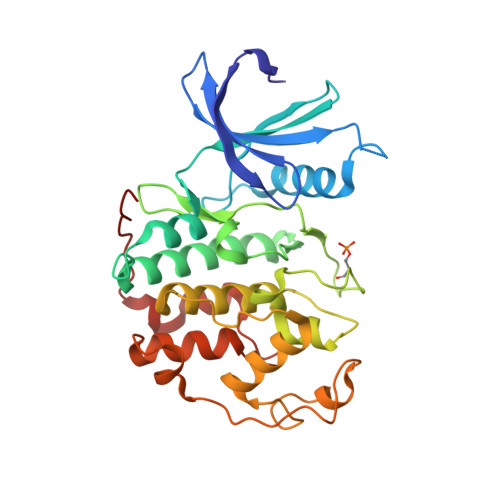
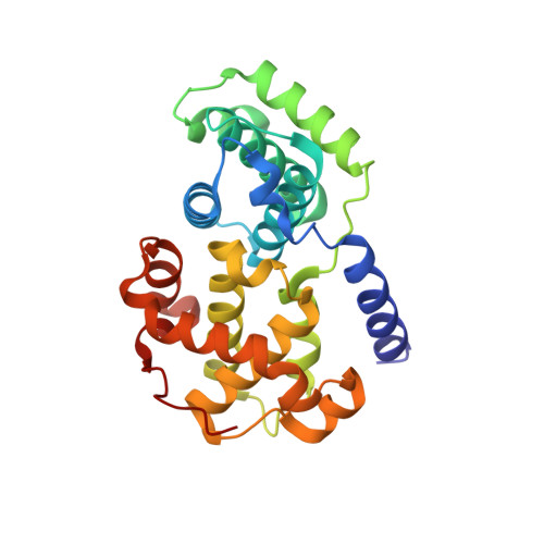
4EOP

Thr 160 phosphorylated CDK2 Q131E - human cyclin A3 complex with the inhibitor RO3306


Experimental Data Snapshot

  • Method: X-RAY DIFFRACTION
  • Resolution: 1.99 Å
  • R-Value Free: 0.241 
  • R-Value Work: 0.216 
  • R-Value Observed: 0.217 

wwPDB Validation   3D Report Full Report


Ligand Structure Quality Assessment 


This is version 1.0 of the entry. See complete history


Literature

An integrated chemical biology approach provides insight into Cdk2 functional redundancy and inhibitor sensitivity.

Echalier, A.Cot, E.Camasses, A.Hodimont, E.Hoh, F.Jay, P.Sheinerman, F.Krasinska, L.Fisher, D.

(2012) Chem Biol 19: 1028-1040

  • DOI: https://doi.org/10.1016/j.chembiol.2012.06.015
  • Primary Citation of Related Structures:  
    4EOI, 4EOJ, 4EOK, 4EOL, 4EOM, 4EON, 4EOO, 4EOP, 4EOQ, 4EOR, 4EOS

  • PubMed Abstract: 

    Cdk2 promotes DNA replication and is a promising cancer therapeutic target, but its functions appear redundant with Cdk1, an essential Cdk affected by most Cdk2 inhibitors. Here, we present an integrated multidisciplinary approach to address Cdk redundancy. Mathematical modeling of enzymology data predicted conditions allowing selective chemical Cdk2 inhibition. Together with experiments in Xenopus egg extracts, this supports a rate-limiting role for Cdk2 in DNA replication. To confirm this we designed inhibitor-resistant (ir)-Cdk2 mutants using a novel bioinformatics approach. Bypassing inhibition with ir-Cdk2 or with Cdk1 shows that Cdk2 is rate-limiting for replication in this system because Cdk1 is insufficiently active. Additionally, crystal structures and kinetics reveal alternative binding modes of Cdk1-selective and Cdk2-selective inhibitors and mechanisms of Cdk2 inhibitor resistance. Our approach thus provides insight into structure, functions, and biochemistry of a cyclin-dependent kinase.


  • Organizational Affiliation

    Université Montpellier I, 34000 Montpellier, France.


Macromolecules
Find similar proteins by:  (by identity cutoff)  |  3D Structure
Entity ID: 1
MoleculeChains Sequence LengthOrganismDetailsImage
Cyclin-dependent kinase 2
A, C
300Homo sapiensMutation(s): 1 
Gene Names: CDK2CDKN2
EC: 2.7.11.22
UniProt & NIH Common Fund Data Resources
Find proteins for P24941 (Homo sapiens)
Explore P24941 
Go to UniProtKB:  P24941
PHAROS:  P24941
GTEx:  ENSG00000123374 
Entity Groups  
Sequence Clusters30% Identity50% Identity70% Identity90% Identity95% Identity100% Identity
UniProt GroupP24941
Sequence Annotations
Expand
  • Reference Sequence
Find similar proteins by:  (by identity cutoff)  |  3D Structure
Entity ID: 2
MoleculeChains Sequence LengthOrganismDetailsImage
Cyclin-A2
B, D
258Homo sapiensMutation(s): 0 
Gene Names: CCNA2CCN1CCNA
UniProt & NIH Common Fund Data Resources
Find proteins for P20248 (Homo sapiens)
Explore P20248 
Go to UniProtKB:  P20248
PHAROS:  P20248
GTEx:  ENSG00000145386 
Entity Groups  
Sequence Clusters30% Identity50% Identity70% Identity90% Identity95% Identity100% Identity
UniProt GroupP20248
Sequence Annotations
Expand
  • Reference Sequence
Small Molecules
Modified Residues  1 Unique
IDChains TypeFormula2D DiagramParent
TPO
Query on TPO
A, C
L-PEPTIDE LINKINGC4 H10 N O6 PTHR
Binding Affinity Annotations 
IDSourceBinding Affinity
1RO PDBBind:  4EOP Ki: 890 (nM) from 1 assay(s)
BindingDB:  4EOP IC50: 253 (nM) from 1 assay(s)
Experimental Data & Validation

Experimental Data

  • Method: X-RAY DIFFRACTION
  • Resolution: 1.99 Å
  • R-Value Free: 0.241 
  • R-Value Work: 0.216 
  • R-Value Observed: 0.217 
  • Space Group: P 21 21 21
Unit Cell:
Length ( Å )Angle ( ˚ )
a = 74.78α = 90
b = 133.39β = 90
c = 147.68γ = 90
Software Package:
Software NamePurpose
ADSCdata collection
MOLREPphasing
REFMACrefinement
MOSFLMdata reduction
SCALAdata scaling

Structure Validation

View Full Validation Report


Ligand Structure Quality Assessment 


Entry History 

Deposition Data

Revision History  (Full details and data files)

  • Version 1.0: 2013-02-06
    Type: Initial release