4EJN | pdb_00004ejn

Crystal structure of autoinhibited form of AKT1 in complex with N-(4-(5-(3-acetamidophenyl)-2-(2-aminopyridin-3-yl)-3H-imidazo[4,5-b]pyridin-3-yl)benzyl)-3-fluorobenzamide


Experimental Data Snapshot

  • Method: X-RAY DIFFRACTION
  • Resolution: 2.19 Å
  • R-Value Free: 
    0.276 (Depositor), 0.279 (DCC) 
  • R-Value Work: 
    0.237 (Depositor), 0.235 (DCC) 
  • R-Value Observed: 
    0.239 (Depositor) 

wwPDB Validation 3D Report Full Report

Validation slider image for 4EJN

Ligand Structure Quality Assessment 


This is version 1.2 of the entry. See complete history

Literature

Discovery and optimization of a series of 3-(3-phenyl-3H-imidazo[4,5-b]pyridin-2-yl)pyridin-2-amines: orally bioavailable, selective, and potent ATP-independent Akt inhibitors.

Ashwell, M.A.Lapierre, J.M.Brassard, C.Bresciano, K.Bull, C.Cornell-Kennon, S.Eathiraj, S.France, D.S.Hall, T.Hill, J.Kelleher, E.Khanapurkar, S.Kizer, D.Koerner, S.Link, J.Liu, Y.Makhija, S.Moussa, M.Namdev, N.Nguyen, K.Nicewonger, R.Palma, R.Szwaya, J.Tandon, M.Uppalapati, U.Vensel, D.Volak, L.P.Volckova, E.Westlund, N.Wu, H.Yang, R.Y.Chan, T.C.

(2012) J Med Chem 55: 5291-5310

  • DOI: https://doi.org/10.1021/jm300276x
  • Primary Citation Related Structures: 
    4EJN

  • PubMed Abstract: 

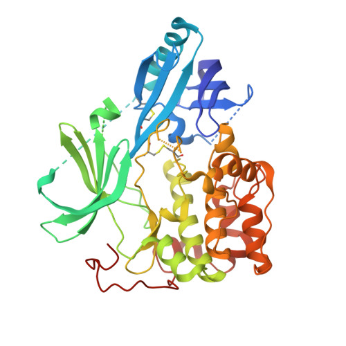
    This paper describes the implementation of a biochemical and biophysical screening strategy to identify and optimize small molecule Akt1 inhibitors that act through a mechanism distinct from that observed for kinase domain ATP-competitive inhibitors. With the aid of an unphosphorylated Akt1 cocrystal structure of 12j solved at 2.25 Å, it was possible to confirm that as a consequence of binding these novel inhibitors, the ATP binding cleft contained a number of hydrophobic residues that occlude ATP binding as expected. These Akt inhibitors potently inhibit intracellular Akt activation and its downstream target (PRAS40) in vitro. In vivo pharmacodynamic and pharmacokinetic studies with two examples, 12e and 12j, showed the series to be similarly effective at inhibiting the activation of Akt and an additional downstream effector (p70S6) following oral dosing in mice.


  • Organizational Affiliation
    • ArQule Inc., 19 Presidential Way, Woburn, Massachusetts 01801, United States.

Macromolecule Content 

  • Total Structure Weight: 52.45 kDa 
  • Atom Count: 3,156 
  • Modeled Residue Count: 376 
  • Deposited Residue Count: 446 
  • Unique protein chains: 1

Macromolecules

Find similar proteins by:|  3D Structure
Entity ID: 1
MoleculeChains  Sequence LengthOrganismDetailsImage
RAC-alpha serine/threonine-protein kinase446Homo sapiensMutation(s): 3 
Gene Names: AKT1PKBRAC
EC: 2.7.11.1
UniProt & NIH Common Fund Data Resources
Find proteins for P31749 (Homo sapiens)
Explore P31749 
Go to UniProtKB:  P31749
PHAROS:  P31749
GTEx:  ENSG00000142208 
Entity Groups
Sequence Clusters30% Identity50% Identity70% Identity90% Identity95% Identity100% Identity
UniProt GroupP31749
Sequence Annotations
Expand
Reference Sequence

Small Molecules

Ligands 3 Unique
IDChains Name / Formula / InChI Key2D Diagram3D Interactions
0R4

Query on 0R4


Download:Ideal Coordinates CCD File
B [auth A]N-(4-{5-[3-(acetylamino)phenyl]-2-(2-aminopyridin-3-yl)-3H-imidazo[4,5-b]pyridin-3-yl}benzyl)-3-fluorobenzamide
C33 H26 F N7 O2
WFDHLKUQFQCHKF-UHFFFAOYSA-N
SBT

Query on SBT


Download:Ideal Coordinates CCD File
C [auth A]2-BUTANOL
C4 H10 O
BTANRVKWQNVYAZ-BYPYZUCNSA-N
EDO

Query on EDO


Download:Ideal Coordinates CCD File
D [auth A]1,2-ETHANEDIOL
C2 H6 O2
LYCAIKOWRPUZTN-UHFFFAOYSA-N

Experimental Data & Validation

Experimental Data

  • Method: X-RAY DIFFRACTION
  • Resolution: 2.19 Å
  • R-Value Free:  0.276 (Depositor), 0.279 (DCC) 
  • R-Value Work:  0.237 (Depositor), 0.235 (DCC) 
  • R-Value Observed: 0.239 (Depositor) 
Space Group: P 21 21 21
Unit Cell:
Length ( Å )Angle ( ˚ )
a = 44.508α = 90
b = 88.214β = 90
c = 126.936γ = 90
Software Package:
Software NamePurpose
ADSCdata collection
PHASERphasing
REFMACrefinement
HKL-2000data reduction
SCALEPACKdata scaling

Structure Validation

View Full Validation Report


Ligand Structure Quality Assessment 


Entry History 

Deposition Data

  • Released Date: 2012-05-23 
  • Deposition Author(s): Eathiraj, S.

Revision History  (Full details and data files)

  • Version 1.0: 2012-05-23
    Type: Initial release
  • Version 1.1: 2013-01-02
    Changes: Database references
  • Version 1.2: 2024-10-16
    Changes: Data collection, Database references, Derived calculations, Structure summary