4EEO | pdb_00004eeo

Crystal structure of human M340H-beta-1,4-galactosyltransferase-1 (M340H-B4GAL-T1) in complex with GLCNAC-BETA1,6-GlcNAc-ALPHA-benzyl


Experimental Data Snapshot

  • Method: X-RAY DIFFRACTION
  • Resolution: 2.30 Å
  • R-Value Free: 
    0.255 (Depositor), 0.249 (DCC) 
  • R-Value Work: 
    0.203 (Depositor) 
  • R-Value Observed: 
    0.205 (Depositor) 

Starting Model: experimental
View more details

wwPDB Validation 3D Report Full Report

Validation slider image for 4EEO

Ligand Structure Quality Assessment 


This is version 2.2 of the entry. See complete history

Literature

Binding of N-acetylglucosamine (GlcNAc) beta 1-6-branched oligosaccharide acceptors to beta 4-galactosyltransferase I reveals a new ligand binding mode.

Ramakrishnan, B.Boeggeman, E.Qasba, P.K.

(2012) J Biological Chem 287: 28666-28674

  • DOI: https://doi.org/10.1074/jbc.M112.373514
  • Primary Citation Related Structures: 
    4EE3, 4EE4, 4EE5, 4EEA, 4EEG, 4EEM, 4EEO

  • PubMed Abstract: 

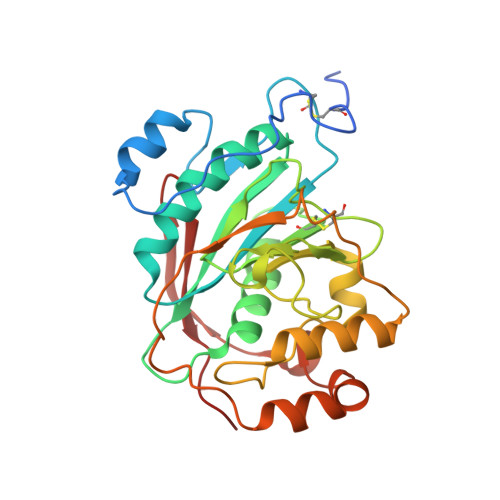
    N-acetyllactosamine is the most prevalent disaccharide moiety in the glycans on the surface of mammalian cells and often found as repeat units in the linear and branched polylactosamines, known as i- and I-antigen, respectively. The β1-4-galactosyltransferase-I (β4Gal-T1) enzyme is responsible for the synthesis of the N-acetyllactosamine moiety. To understand its oligosaccharide acceptor specificity, we have previously investigated the binding of tri- and pentasaccharides of N-glycan with a GlcNAc at their nonreducing end and found that the extended sugar moiety in these acceptor substrates binds to the crevice present at the acceptor substrate binding site of the β4Gal-T1 molecule. Here we report seven crystal structures of β4Gal-T1 in complex with an oligosaccharide acceptor with a nonreducing end GlcNAc that has a β1-6-glycosidic link and that are analogous to either N-glycan or i/I-antigen. In the crystal structure of the complex of β4Gal-T1 with I-antigen analog pentasaccharide, the β1-6-branched GlcNAc moiety is bound to the sugar acceptor binding site of the β4Gal-T1 molecule in a way similar to the crystal structures described previously; however, the extended linear tetrasaccharide moiety does not interact with the previously found extended sugar binding site on the β4Gal-T1 molecule. Instead, it interacts with the different hydrophobic surface of the protein molecule formed by the residues Tyr-276, Trp-310, and Phe-356. Results from the present and previous studies suggest that β4Gal-T1 molecule has two different oligosaccharide binding regions for the binding of the extended oligosaccharide moiety of the acceptor substrate.


  • Organizational Affiliation
    • Structural Glycobiology Section, SAIC-Frederick, Inc., Center for Cancer Research Nanobiology Program, Center for Cancer Research, Frederick National Laboratory for Cancer Research, National Institutes of Health, Frederick, Maryland 21702, USA.

Macromolecule Content 

  • Total Structure Weight: 102.57 kDa 
  • Atom Count: 7,408 
  • Modeled Residue Count: 819 
  • Deposited Residue Count: 861 
  • Unique protein chains: 1

Macromolecules

Find similar proteins by:|  3D Structure
Entity ID: 1
MoleculeChains  Sequence LengthOrganismDetailsImage
Beta-1,4-galactosyltransferase 1
A, B, C
287Homo sapiensMutation(s): 3 
Gene Names: B4GALT1GGTB2
EC: 2.4.1 (PDB Primary Data), 2.4.1.22 (PDB Primary Data), 2.4.1.90 (PDB Primary Data), 2.4.1.38 (PDB Primary Data), 2.4.1.275 (UniProt)
UniProt & NIH Common Fund Data Resources
Find proteins for P15291 (Homo sapiens)
Explore P15291 
Go to UniProtKB:  P15291
PHAROS:  P15291
GTEx:  ENSG00000086062 
Entity Groups
Sequence Clusters30% Identity50% Identity70% Identity90% Identity95% Identity100% Identity
UniProt GroupP15291
Sequence Annotations
Expand
Reference Sequence

Oligosaccharides

Help  
Entity ID: 2
MoleculeChains Length2D Diagram GlycosylationD Interactions
2-acetamido-2-deoxy-beta-D-glucopyranose-(1-6)-benzyl 2-acetamido-2-deoxy-alpha-D-glucopyranoside
D, E, F
2N/AN/A
Glycosylation Resources
GlyTouCan: G86816WG
GlyCosmos: G86816WG

Small Molecules

Ligands 4 Unique
IDChains Name / Formula / InChI Key2D Diagram3D Interactions
UDP

Query on UDP


Download:Ideal Coordinates CCD File
G [auth A],
N [auth B],
V [auth C]
URIDINE-5'-DIPHOSPHATE
C9 H14 N2 O12 P2
XCCTYIAWTASOJW-XVFCMESISA-N
SO4

Query on SO4


Download:Ideal Coordinates CCD File
I [auth A]
J [auth A]
K [auth A]
L [auth A]
P [auth B]
I [auth A],
J [auth A],
K [auth A],
L [auth A],
P [auth B],
Q [auth B],
R [auth B],
S [auth B],
T [auth B],
X [auth C],
Y [auth C]
SULFATE ION
O4 S
QAOWNCQODCNURD-UHFFFAOYSA-L
GOL

Query on GOL


Download:Ideal Coordinates CCD File
M [auth A],
U [auth B],
Z [auth C]
GLYCEROL
C3 H8 O3
PEDCQBHIVMGVHV-UHFFFAOYSA-N
MN

Query on MN


Download:Ideal Coordinates CCD File
H [auth A],
O [auth B],
W [auth C]
MANGANESE (II) ION
Mn
WAEMQWOKJMHJLA-UHFFFAOYSA-N

Experimental Data & Validation

Experimental Data

  • Method: X-RAY DIFFRACTION
  • Resolution: 2.30 Å
  • R-Value Free:  0.255 (Depositor), 0.249 (DCC) 
  • R-Value Work:  0.203 (Depositor) 
  • R-Value Observed: 0.205 (Depositor) 
Space Group: C 2 2 21
Unit Cell:
Length ( Å )Angle ( ˚ )
a = 107.804α = 90
b = 198.217β = 90
c = 143.732γ = 90
Software Package:
Software NamePurpose
HKL-2000data collection
REFMACrefinement
HKL-2000data reduction
HKL-2000data scaling

Structure Validation

View Full Validation Report


Ligand Structure Quality Assessment 


Entry History 

Deposition Data

Revision History  (Full details and data files)

  • Version 1.0: 2012-07-04
    Type: Initial release
  • Version 1.1: 2014-03-26
    Changes: Database references
  • Version 1.2: 2017-11-15
    Changes: Refinement description
  • Version 2.0: 2020-07-29
    Type: Remediation
    Reason: Carbohydrate remediation
    Changes: Atomic model, Data collection, Database references, Derived calculations, Structure summary
  • Version 2.1: 2023-09-13
    Changes: Data collection, Database references, Refinement description, Structure summary
  • Version 2.2: 2024-11-20
    Changes: Structure summary