4E0K

Crystal Structure of the amyloid-fibril forming peptide KDWSFY derived from human Beta 2 Microglobulin (58-63)


Experimental Data Snapshot

  • Method: X-RAY DIFFRACTION
  • Resolution: 0.97 Å
  • R-Value Free: 0.131 
  • R-Value Work: 0.112 
  • R-Value Observed: 0.114 

wwPDB Validation   3D Report Full Report


This is version 1.2 of the entry. See complete history


Literature

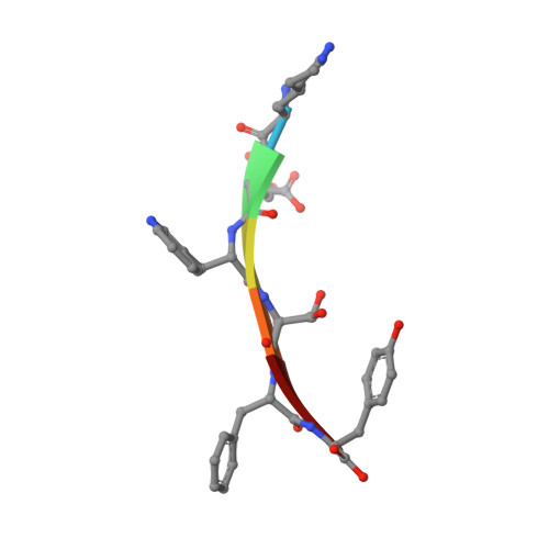
Out-of-register beta-sheets suggest a pathway to toxic amyloid aggregates.

Liu, C.Zhao, M.Jiang, L.Cheng, P.N.Park, J.Sawaya, M.R.Pensalfini, A.Gou, D.Berk, A.J.Glabe, C.G.Nowick, J.Eisenberg, D.

(2012) Proc Natl Acad Sci U S A 109: 20913-20918

  • DOI: https://doi.org/10.1073/pnas.1218792109
  • Primary Citation of Related Structures:  
    4E0K, 4E0L, 4E0M, 4E0N, 4E0O

  • PubMed Abstract: 

    Although aberrant protein aggregation has been conclusively linked to dozens of devastating amyloid diseases, scientists remain puzzled about the molecular features that render amyloid fibrils or small oligomers toxic. Here, we report a previously unobserved type of amyloid fibril that tests as cytotoxic: one in which the strands of the contributing β-sheets are out of register. In all amyloid fibrils previously characterized at the molecular level, only in-register β-sheets have been observed, in which each strand makes its full complement of hydrogen bonds with the strands above and below it in the fibril. In out-of-register sheets, strands are sheared relative to one another, leaving dangling hydrogen bonds. Based on this finding, we designed out-of-register β-sheet amyloid mimics, which form both cylindrin-like oligomers and fibrils, and these mimics are cytotoxic. Structural and energetic considerations suggest that out-of-register fibrils can readily convert to toxic cylindrins. We propose that out-of-register β-sheets and their related cylindrins are part of a toxic amyloid pathway, which is distinct from the more energetically favored in-register amyloid pathway.


  • Organizational Affiliation

    UCLA-DOE Institute for Genomics and Proteomics, The Howard Hughes Medical Institute, Molecular Biology Institute and Molecular Biology Institute, University of California, Los Angeles, CA 90095, USA.


Macromolecules

Find similar proteins by:  Sequence   |   3D Structure  

Entity ID: 1
MoleculeChains Sequence LengthOrganismDetailsImage
Amyloidogenic peptide segment KDWSFY
A, B, C, D
6N/AMutation(s): 0 
UniProt & NIH Common Fund Data Resources
Find proteins for P61769 (Homo sapiens)
Explore P61769 
Go to UniProtKB:  P61769
PHAROS:  P61769
GTEx:  ENSG00000166710 
Entity Groups  
UniProt GroupP61769
Sequence Annotations
Expand
  • Reference Sequence
Small Molecules
Ligands 2 Unique
IDChains Name / Formula / InChI Key2D Diagram3D Interactions
PE4
Query on PE4

Download Ideal Coordinates CCD File 
E [auth B],
G [auth C]
2-{2-[2-(2-{2-[2-(2-ETHOXY-ETHOXY)-ETHOXY]-ETHOXY}-ETHOXY)-ETHOXY]-ETHOXY}-ETHANOL
C16 H34 O8
PJWQOENWHPEPKI-UHFFFAOYSA-N
NA
Query on NA

Download Ideal Coordinates CCD File 
F [auth B]SODIUM ION
Na
FKNQFGJONOIPTF-UHFFFAOYSA-N
Experimental Data & Validation

Experimental Data

  • Method: X-RAY DIFFRACTION
  • Resolution: 0.97 Å
  • R-Value Free: 0.131 
  • R-Value Work: 0.112 
  • R-Value Observed: 0.114 
  • Space Group: P 61
Unit Cell:
Length ( Å )Angle ( ˚ )
a = 62.37α = 90
b = 62.37β = 90
c = 11.73γ = 120
Software Package:
Software NamePurpose
XSCALEdata scaling
PHENIXrefinement
PDB_EXTRACTdata extraction

Structure Validation

View Full Validation Report


Entry History 

Deposition Data

Revision History  (Full details and data files)

  • Version 1.0: 2012-12-19
    Type: Initial release
  • Version 1.1: 2013-06-19
    Changes: Database references
  • Version 1.2: 2024-02-28
    Changes: Data collection, Database references, Derived calculations