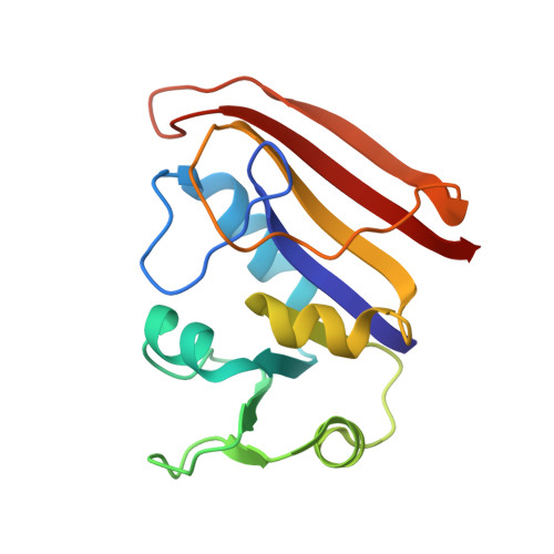
4DFR

CRYSTAL STRUCTURES OF ESCHERICHIA COLI AND LACTOBACILLUS CASEI DIHYDROFOLATE REDUCTASE REFINED AT 1.7 ANGSTROMS RESOLUTION. I. GENERAL FEATURES AND BINDING OF METHOTREXATE


Experimental Data Snapshot

  • Method: X-RAY DIFFRACTION
  • Resolution: 1.70 Å
  • R-Value Work: 0.155 

wwPDB Validation   3D Report Full Report


This is version 1.3 of the entry. See complete history


Literature

Crystal structures of Escherichia coli and Lactobacillus casei dihydrofolate reductase refined at 1.7 A resolution. I. General features and binding of methotrexate.

Bolin, J.T.Filman, D.J.Matthews, D.A.Hamlin, R.C.Kraut, J.

(1982) J Biol Chem 257: 13650-13662

  • Primary Citation of Related Structures:  
    3DFR, 4DFR

  • PubMed Abstract: 
  • X-ray data have been extended to 1.7 A for a binary complex of Escherichia coli dihydrofolate reductase with methotrexate and a ternary complex of Lactobacillus casei dihydrofolate reductase with methotrexate and NADPH. Models for both structures have been refined to R factors of 0 ...

    X-ray data have been extended to 1.7 A for a binary complex of Escherichia coli dihydrofolate reductase with methotrexate and a ternary complex of Lactobacillus casei dihydrofolate reductase with methotrexate and NADPH. Models for both structures have been refined to R factors of 0.15 and include parameters for fixed and liquid solvent. The two species of dihydrofolate reductase resemble one another even more closely than was thought to be the case prior to refinement. Several new structural features have also been discovered. Among them are a cis peptide linking Gly-97 and Gly-98 (L. Casei numbering) in both species, an alpha helix involving residues 43 through 50 in the E. coli enzyme, and the existence of what may be a specific hydration site on exposed alpha helices. Refinement has led to a revised description of the details of methotrexate binding. We now see that a fixed water molecule mediates the interaction between methotrexate's 2-amino group and Thr-116 (L. casei numbering) and that the inhibitor's 4-amino group makes two hydrogen bonds with the enzyme (instead of one). Other revisions are also discussed. A hypothetical model for substrate binding is proposed in which the pteridine ring is turned upside down while all protein and solvent atoms remain fixed. Asp-26 in this model is hydrogen bonded to the substrate's 2-amino group and to N3.


    Related Citations: 
    • Effect of Single Amino Acid Replacements on the Folding and Stability of Dihydrofolate Reductase from Escherichia Coli
      Perry, K.M., Onuffer, J.J., Touchette, N.A., Herndon, C.S., Gittelman, M.S., Matthews, C.R., Chen, J.-T., Mayer, R.J., Taira, K., Benkovic, S.J., Howell, E.E., Kraut, J.
      (1987) Biochemistry 26: 2674
    • Crystal Structures of Escherichia Coli and Lactobacillus Casei Dihydrofolate Reductase Refined at 1.7 Angstroms Resolution. II. Environment of Bound Nadph and Implications for Catalysis
      Filman, D.J., Bolin, J.T., Matthews, D.A., Kraut, J.
      (1982) J Biol Chem 257: 13663
    • Crystal Structure of Avian Dihydrofolate Reductase Containing Phenyltriazine and Nadph
      Volz, K.W., Matthews, D.A., Alden, R.A., Freer, S.T., Hansch, C., Kaufman, B.T., Kraut, J.
      (1982) J Biol Chem 257: 2528
    • Interpretation of Nuclear Magnetic Resonance Spectra for Lactobacillus Casei Dihydrofolate Reductase Based on the X-Ray Structure of the Enzyme-Methotrexate-Nadph Complex
      Matthews, D.A.
      (1979) Biochemistry 18: 1602
    • Dihydrofolate Reductase from Lactobacillus Casei. Stereochemistry of Nadph Binding
      Matthews, D.A., Alden, R.A., Freer, S.T., Xuong, N.-H., Kraut, J.
      (1979) J Biol Chem 254: 4144
    • Proton Magnetic Resonance Studies on Escherichia Coli Dihydrofolate Reductase. Assignment of Histidine C-2 Protons in Binary Complexes with Folates on the Basis of the Crystal Structure with Methotrexate and on Chemical Modifications
      Poe, M., Hoogsteen, K., Matthews, D.A.
      (1979) J Biol Chem 254: 8143
    • Dihydrofolate Reductase from Lactobacillus Casei. X-Ray Structure of the Enzyme-Methotrexate-Nadph Complex
      Matthews, D.A., Alden, R.A., Bolin, J.T., Filman, D.J., Freer, S.T., Hamlin, R., Hol, W.G.J., Kisliuk, R.L., Pastore, E.J., Plante, L.T., Xuong, N.-H., Kraut, J.
      (1978) J Biol Chem 253: 6946
    • Dihydrofolate Reductase. The Amino Acid Sequence of the Enzyme from a Methotrexate-Resistant Mutant of Escherichia Coli
      Bennett, C.D., Rodkey, J.A., Sondey, J.M., Hirschmann, R.
      (1978) Biochemistry 17: 1328
    • Dihydrofolate Reductase. X-Ray Structure of the Binary Complex with Methotrexate
      Matthews, D.A., Alden, R.A., Bolin, J.T., Freer, S.T., Hamlin, R., Xuong, N., Kraut, J., Poe, M., Williams, M., Hoogsteen, K.
      (1977) Science 197: 452
    • Dihydrofolate Reductase. Purification and Characterization of the Enzyme from an Amethopterin-Resistant Mutant of Escherichia Coli
      Poe, M., Greenfield, N.J., Hirshfield, J.M., Williams, M.N., Hoogsteen, K.
      (1972) Biochemistry 11: 1023

    Organizational Affiliation

    School of Molecular and Microbial Sciences, University of Queensland, Brisbane 4072, Australia.


Macromolecules
Find similar proteins by:  (by identity cutoff)  |  3D Structure
Entity ID: 1
MoleculeChainsSequence LengthOrganismDetailsImage
DIHYDROFOLATE REDUCTASE
A, B
159Escherichia coli BMutation(s): 0 
EC: 1.5.1.3
UniProt
Find proteins for P0ABQ4 (Escherichia coli (strain K12))
Explore P0ABQ4 
Go to UniProtKB:  P0ABQ4
Entity Groups  
Sequence Clusters30% Identity50% Identity70% Identity90% Identity95% Identity100% Identity
UniProt GroupP0ABQ4
Protein Feature View
Expand
  • Reference Sequence
Small Molecules
Ligands 3 Unique
IDChainsName / Formula / InChI Key2D Diagram3D Interactions
MTX
Query on MTX

Download Ideal Coordinates CCD File 
D [auth A],
G [auth B]
METHOTREXATE
C20 H22 N8 O5
FBOZXECLQNJBKD-ZDUSSCGKSA-N
 Ligand Interaction
CA
Query on CA

Download Ideal Coordinates CCD File 
F [auth B]CALCIUM ION
Ca
BHPQYMZQTOCNFJ-UHFFFAOYSA-N
 Ligand Interaction
CL
Query on CL

Download Ideal Coordinates CCD File 
C [auth A],
E [auth B]
CHLORIDE ION
Cl
VEXZGXHMUGYJMC-UHFFFAOYSA-M
 Ligand Interaction
Binding Affinity Annotations 
IDSourceBinding Affinity
MTX BindingDB:  4DFR IC50: min: 3, max: 8.8 (nM) from 3 assay(s)
EC50: 1 (nM) from 1 assay(s)
Experimental Data & Validation

Experimental Data

  • Method: X-RAY DIFFRACTION
  • Resolution: 1.70 Å
  • R-Value Work: 0.155 
  • Space Group: P 61
Unit Cell:
Length ( Å )Angle ( ˚ )
a = 93.22α = 90
b = 93.22β = 90
c = 73.56γ = 120

Structure Validation

View Full Validation Report


Entry History 

Deposition Data

Revision History  (Full details and data files)

  • Version 1.0: 1982-10-21
    Type: Initial release
  • Version 1.1: 2008-03-03
    Changes: Version format compliance
  • Version 1.2: 2011-07-13
    Changes: Source and taxonomy, Version format compliance
  • Version 1.3: 2017-11-29
    Changes: Derived calculations, Other, Structure summary