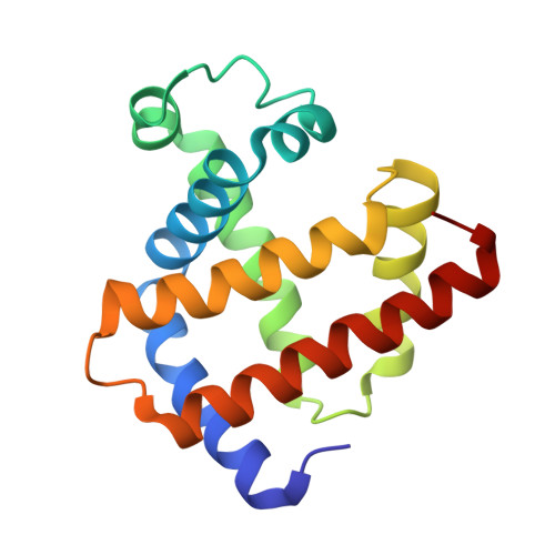
Towards protein-crystal centering using second-harmonic generation (SHG) microscopy.
Kissick, D.J., Dettmar, C.M., Becker, M., Mulichak, A.M., Cherezov, V., Ginell, S.L., Battaile, K.P., Keefe, L.J., Fischetti, R.F., Simpson, G.J.(2013) Acta Crystallogr D Biol Crystallogr 69: 843-851
- PubMed: 23633594
- DOI: https://doi.org/10.1107/S0907444913002746
- Primary Citation of Related Structures:
4DC5, 4DC6, 4DC7, 4DC8 - PubMed Abstract:
The potential of second-harmonic generation (SHG) microscopy for automated crystal centering to guide synchrotron X-ray diffraction of protein crystals was explored. These studies included (i) comparison of microcrystal positions in cryoloops as determined by SHG imaging and by X-ray diffraction rastering and (ii) X-ray structure determinations of selected proteins to investigate the potential for laser-induced damage from SHG imaging. In studies using β2 adrenergic receptor membrane-protein crystals prepared in lipidic mesophase, the crystal locations identified by SHG images obtained in transmission mode were found to correlate well with the crystal locations identified by raster scanning using an X-ray minibeam. SHG imaging was found to provide about 2 µm spatial resolution and shorter image-acquisition times. The general insensitivity of SHG images to optical scatter enabled the reliable identification of microcrystals within opaque cryocooled lipidic mesophases that were not identified by conventional bright-field imaging. The potential impact of extended exposure of protein crystals to five times a typical imaging dose from an ultrafast laser source was also assessed. Measurements of myoglobin and thaumatin crystals resulted in no statistically significant differences between structures obtained from diffraction data acquired from exposed and unexposed regions of single crystals. Practical constraints for integrating SHG imaging into an active beamline for routine automated crystal centering are discussed.
- Department of Chemistry, Purdue University, West Lafayette, IN 47907, USA.
Organizational Affiliation:


