Three-Dimensional Electron Microscopy Reconstruction and Cysteine-Mediated Crosslinking Provide a Model of the T3Ss Needle Tip Complex.
Cheung, M., Shen, D., Makino, F., Kato, T., Roehrich, A.D., Martinez-Argudo, I., Walker, M.L., Murillo, I., Liu, X., Pain, M., Brown, J., Frazer, G., Mantell, J., Mina, P., Todd, T., Sessions, R.B., Namba, K., Blocker, A.J.(2015) Mol Microbiol 95: 31
- PubMed: 25353930 Search on PubMedSearch on PubMed Central
- DOI: https://doi.org/10.1111/mmi.12843
- Primary Citation Related Structures:
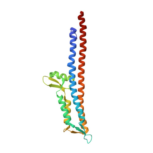
4D3E - PubMed Abstract:
Type III secretion systems are found in many Gram-negative bacteria. They are activated by contact with eukaryotic cells and inject virulence proteins inside them. Host cell detection requires a protein complex located at the tip of the device's external injection needle. The Shigella tip complex (TC) is composed of IpaD, a hydrophilic protein, and IpaB, a hydrophobic protein, which later forms part of the injection pore in the host membrane. Here we used labelling and crosslinking methods to show that TCs from a ΔipaB strain contain five IpaD subunits while the TCs from wild-type can also contain one IpaB and four IpaD subunits. Electron microscopy followed by single particle and helical image analysis was used to reconstruct three-dimensional images of TCs at ∼ 20 Å resolution. Docking of an IpaD crystal structure, constrained by the crosslinks observed, reveals that TC organisation is different from that of all previously proposed models. Our findings suggest new mechanisms for TC assembly and function. The TC is the only site within these secretion systems targeted by disease-protecting antibodies. By suggesting how these act, our work will allow improvement of prophylactic and therapeutic strategies.
- Schools of Cellular & Molecular Medicine and Biochemistry, University of Bristol, Bristol, BS8 1TD, UK.
Organizational Affiliation: