4D2G

Crystal structure of human PCNA in complex with p15 peptide


Experimental Data Snapshot

  • Method: X-RAY DIFFRACTION
  • Resolution: 2.65 Å
  • R-Value Free: 0.250 
  • R-Value Work: 0.170 
  • R-Value Observed: 0.174 

wwPDB Validation   3D Report Full Report


This is version 1.2 of the entry. See complete history


Literature

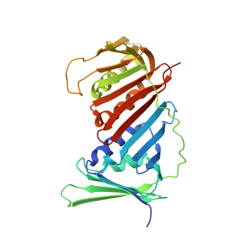
Structure of P15(Paf)-PCNA Complex and Implications for Clamp Sliding During DNA Replication and Repair.

De Biasio, A.De Opakua, A.I.Mortuza, G.B.Molina, R.Cordeiro, T.N.Castillo, F.Villate, M.Merino, N.Delgado, S.Gil-Carton, D.Luque, I.Diercks, T.Bernado, P.Montoya, G.Blanco, F.J.

(2015) Nat Commun 6: 6439

  • DOI: https://doi.org/10.1038/ncomms7439
  • Primary Citation of Related Structures:  
    4D2G

  • PubMed Abstract: 

    The intrinsically disordered protein p15(PAF) regulates DNA replication and repair by binding to the proliferating cell nuclear antigen (PCNA) sliding clamp. We present the structure of the human p15(PAF)-PCNA complex. Crystallography and NMR show the central PCNA-interacting protein motif (PIP-box) of p15(PAF) tightly bound to the front-face of PCNA. In contrast to other PCNA-interacting proteins, p15(PAF) also contacts the inside of, and passes through, the PCNA ring. The disordered p15(PAF) termini emerge at opposite faces of the ring, but remain protected from 20S proteasomal degradation. Both free and PCNA-bound p15(PAF) binds DNA mainly through its histone-like N-terminal tail, while PCNA does not, and a model of the ternary complex with DNA inside the PCNA ring is consistent with electron micrographs. We propose that p15(PAF) acts as a flexible drag that regulates PCNA sliding along the DNA and facilitates the switch from replicative to translesion synthesis polymerase binding.


  • Organizational Affiliation

    Structural Biology Unit, CIC bioGUNE, Parque Tecnológico de Bizkaia Edificio 800, 48160 Derio, Spain.


Macromolecules
Find similar proteins by:  (by identity cutoff)  |  3D Structure
Entity ID: 1
MoleculeChains Sequence LengthOrganismDetailsImage
PROLIFERATING CELL NUCLEAR ANTIGEN
A, B, C
264Homo sapiensMutation(s): 0 
UniProt & NIH Common Fund Data Resources
Find proteins for P12004 (Homo sapiens)
Explore P12004 
Go to UniProtKB:  P12004
PHAROS:  P12004
GTEx:  ENSG00000132646 
Entity Groups  
Sequence Clusters30% Identity50% Identity70% Identity90% Identity95% Identity100% Identity
UniProt GroupP12004
Sequence Annotations
Expand
  • Reference Sequence

Find similar proteins by:  Sequence   |   3D Structure  

Entity ID: 2
MoleculeChains Sequence LengthOrganismDetailsImage
P15
D, E
21synthetic constructMutation(s): 0 
UniProt & NIH Common Fund Data Resources
Find proteins for Q15004 (Homo sapiens)
Explore Q15004 
Go to UniProtKB:  Q15004
PHAROS:  Q15004
GTEx:  ENSG00000166803 
Entity Groups  
Sequence Clusters30% Identity50% Identity70% Identity90% Identity95% Identity100% Identity
UniProt GroupQ15004
Sequence Annotations
Expand
  • Reference Sequence
Experimental Data & Validation

Experimental Data

  • Method: X-RAY DIFFRACTION
  • Resolution: 2.65 Å
  • R-Value Free: 0.250 
  • R-Value Work: 0.170 
  • R-Value Observed: 0.174 
  • Space Group: P 1 21 1
Unit Cell:
Length ( Å )Angle ( ˚ )
a = 76.053α = 90
b = 41.228β = 105.39
c = 141.121γ = 90
Software Package:
Software NamePurpose
PHENIXrefinement
XDSdata reduction
SCALAdata scaling
PHASERphasing

Structure Validation

View Full Validation Report


Entry History 

Revision History  (Full details and data files)

  • Version 1.0: 2015-03-18
    Type: Initial release
  • Version 1.1: 2015-03-25
    Changes: Database references
  • Version 1.2: 2023-12-20
    Changes: Data collection, Database references, Other, Refinement description