4CO6

Crystal structure of the Nipah virus RNA free nucleoprotein- phosphoprotein complex


Experimental Data Snapshot

  • Method: X-RAY DIFFRACTION
  • Resolution: 2.50 Å
  • R-Value Free: 0.259 
  • R-Value Work: 0.192 
  • R-Value Observed: 0.195 

wwPDB Validation   3D Report Full Report


This is version 1.2 of the entry. See complete history


Literature

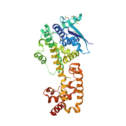
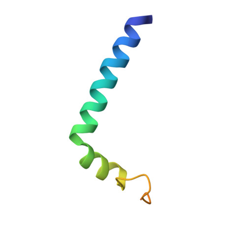
Structure of Nipah Virus Unassembled Nucleoprotein in Complex with its Viral Chaperone.

Yabukarksi, F.Lawrence, P.Tarbouriech, N.Bourhis, J.M.Delaforge, E.Jensen, M.R.Ruigrok, R.W.H.Blackledge, M.Volchkov, V.Jamin, M.

(2014) Nat Struct Mol Biol 21: 754

  • DOI: https://doi.org/10.1038/nsmb.2868
  • Primary Citation of Related Structures:  
    4CO6

  • PubMed Abstract: 

    Nipah virus (NiV) is a highly pathogenic emergent paramyxovirus causing deadly encephalitis in humans. Its replication requires a constant supply of unassembled nucleoprotein (N(0)) in complex with its viral chaperone, the phosphoprotein (P). To elucidate the chaperone function of P, we reconstituted NiV the N(0)-P core complex and determined its crystal structure. The binding of the N-terminal region of P blocks the polymerization of N by interfering with subdomain exchange between N protomers and keeps N(0) in an open conformation, ready to grasp an RNA molecule. We found that a peptide derived from the N-binding region of P protects cells against viral infection and demonstrated by structure-based mutagenesis that this peptide acts by inhibiting N(0)-P formation. These results provide new insights about the assembly of N along genomic RNA and validate the N(0)-P complex as a target for drug development.


  • Organizational Affiliation

    1] Université Grenoble Alpes, Unit of Virus Host Cell Interactions, Grenoble, France. [2] CNRS, Unit of Virus Host Cell Interactions, Grenoble, France.


Macromolecules
Find similar proteins by:  (by identity cutoff)  |  3D Structure
Entity ID: 1
MoleculeChains Sequence LengthOrganismDetailsImage
NUCLEOPROTEIN
A, B, C
356Henipavirus nipahenseMutation(s): 0 
UniProt
Find proteins for Q9IK92 (Nipah virus)
Explore Q9IK92 
Go to UniProtKB:  Q9IK92
Entity Groups  
Sequence Clusters30% Identity50% Identity70% Identity90% Identity95% Identity100% Identity
UniProt GroupQ9IK92
Sequence Annotations
Expand
  • Reference Sequence
Find similar proteins by:  (by identity cutoff)  |  3D Structure
Entity ID: 2
MoleculeChains Sequence LengthOrganismDetailsImage
PHOSPHOPROTEIN
D, E, F
52Henipavirus nipahenseMutation(s): 0 
UniProt
Find proteins for Q9IK91 (Nipah virus)
Explore Q9IK91 
Go to UniProtKB:  Q9IK91
Entity Groups  
Sequence Clusters30% Identity50% Identity70% Identity90% Identity95% Identity100% Identity
UniProt GroupQ9IK91
Sequence Annotations
Expand
  • Reference Sequence
Small Molecules
Modified Residues  1 Unique
IDChains TypeFormula2D DiagramParent
MSE
Query on MSE
A, B, C
L-PEPTIDE LINKINGC5 H11 N O2 SeMET
Experimental Data & Validation

Experimental Data

  • Method: X-RAY DIFFRACTION
  • Resolution: 2.50 Å
  • R-Value Free: 0.259 
  • R-Value Work: 0.192 
  • R-Value Observed: 0.195 
  • Space Group: P 21 21 21
Unit Cell:
Length ( Å )Angle ( ˚ )
a = 82.89α = 90
b = 98.96β = 90
c = 156.91γ = 90
Software Package:
Software NamePurpose
PHENIXrefinement
XDSdata reduction
XDSdata scaling
HKL2MAPphasing

Structure Validation

View Full Validation Report


Entry History 

Deposition Data

Revision History  (Full details and data files)

  • Version 1.0: 2014-08-13
    Type: Initial release
  • Version 1.1: 2014-08-20
    Changes: Database references
  • Version 1.2: 2014-09-17
    Changes: Database references