4CGY | pdb_00004cgy

Crystal structure of the human topoisomerase III alpha-RMI1 complex


Experimental Data Snapshot

  • Method: X-RAY DIFFRACTION
  • Resolution: 2.85 Å
  • R-Value Free: 
    0.231 (Depositor), 0.232 (DCC) 
  • R-Value Work: 
    0.197 (Depositor), 0.203 (DCC) 
  • R-Value Observed: 
    0.199 (Depositor) 

Starting Models: experimental
View more details

wwPDB Validation 3D Report Full Report

Validation slider image for 4CGY

This is version 1.4 of the entry. See complete history

Literature

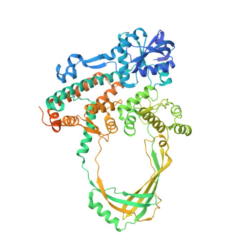
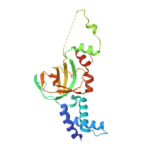
Structural and Mechanistic Insight Into Holliday-Junction Dissolution by Topoisomerase Iiialpha and Rmi1

Bocquet, N.Bizard, A.H.Abdulrahman, W.Larsen, N.B.Faty, M.Cavadini, S.Bunker, R.D.Kowalczykowski, S.C.Cejka, P.Hickson, I.D.Thoma, N.H.

(2014) Nat Struct Mol Biol 21: 261

  • DOI: https://doi.org/10.1038/nsmb.2775
  • Primary Citation Related Structures: 
    4CGY, 4CHT

  • PubMed Abstract: 

    Repair of DNA double-strand breaks via homologous recombination can produce double Holliday junctions (dHJs) that require enzymatic separation. Topoisomerase IIIα (TopIIIα) together with RMI1 disentangles the final hemicatenane intermediate obtained once dHJs have converged. How binding of RMI1 to TopIIIα influences it to behave as a hemicatenane dissolvase, rather than as an enzyme that relaxes DNA topology, is unknown. Here, we present the crystal structure of human TopIIIα complexed to the first oligonucleotide-binding domain (OB fold) of RMI1. TopIII assumes a toroidal type 1A topoisomerase fold. RMI1 attaches to the edge of the gate in TopIIIα through which DNA passes. RMI1 projects a 23-residue loop into the TopIIIα gate, thereby influencing the dynamics of its opening and closing. Our results provide a mechanistic rationale for how RMI1 stabilizes TopIIIα-gate opening to enable dissolution and illustrate how binding partners modulate topoisomerase function.


  • Organizational Affiliation
    • Friedrich Miescher Institute for Biomedical Research, Basel, Switzerland.

Macromolecule Content 

  • Total Structure Weight: 111.25 kDa 
  • Atom Count: 6,602 
  • Modeled Residue Count: 824 
  • Deposited Residue Count: 973 
  • Unique protein chains: 2

Macromolecules

Find similar proteins by:|  3D Structure
Entity ID: 1
MoleculeChains  Sequence LengthOrganismDetailsImage
DNA TOPOISOMERASE 3-ALPHA754Homo sapiensMutation(s): 0 
EC: 5.99.1.2 (PDB Primary Data), 5.6.2.1 (UniProt)
UniProt & NIH Common Fund Data Resources
Find proteins for Q13472 (Homo sapiens)
Explore Q13472 
Go to UniProtKB:  Q13472
PHAROS:  Q13472
GTEx:  ENSG00000177302 
Entity Groups
Sequence Clusters30% Identity50% Identity70% Identity90% Identity95% Identity100% Identity
UniProt GroupQ13472
Sequence Annotations
Expand
Reference Sequence
Find similar proteins by:|  3D Structure
Entity ID: 2
MoleculeChains  Sequence LengthOrganismDetailsImage
RECQ-MEDIATED GENOME INSTABILITY PROTEIN 1219Homo sapiensMutation(s): 0 
UniProt & NIH Common Fund Data Resources
Find proteins for Q9H9A7 (Homo sapiens)
Explore Q9H9A7 
Go to UniProtKB:  Q9H9A7
PHAROS:  Q9H9A7
GTEx:  ENSG00000178966 
Entity Groups
Sequence Clusters30% Identity50% Identity70% Identity90% Identity95% Identity100% Identity
UniProt GroupQ9H9A7
Sequence Annotations
Expand
Reference Sequence

Small Molecules

Ligands 1 Unique
IDChains Name / Formula / InChI Key2D Diagram3D Interactions
MG

Query on MG


Download:Ideal Coordinates CCD File
C [auth A]MAGNESIUM ION
Mg
JLVVSXFLKOJNIY-UHFFFAOYSA-N

Experimental Data & Validation

Experimental Data

  • Method: X-RAY DIFFRACTION
  • Resolution: 2.85 Å
  • R-Value Free:  0.231 (Depositor), 0.232 (DCC) 
  • R-Value Work:  0.197 (Depositor), 0.203 (DCC) 
  • R-Value Observed: 0.199 (Depositor) 
Space Group: P 41 21 2
Unit Cell:
Length ( Å )Angle ( ˚ )
a = 94.01α = 90
b = 94.01β = 90
c = 381.69γ = 90
Software Package:
Software NamePurpose
BUSTERrefinement
XDSdata reduction
XDSdata scaling
PHASERphasing

Structure Validation

View Full Validation Report


Entry History 

Deposition Data

Revision History  (Full details and data files)

  • Version 1.0: 2014-02-12
    Type: Initial release
  • Version 1.1: 2014-02-19
    Changes: Database references
  • Version 1.2: 2014-03-19
    Changes: Database references
  • Version 1.3: 2019-04-03
    Changes: Data collection, Other, Source and taxonomy
  • Version 1.4: 2023-12-20
    Changes: Data collection, Database references, Derived calculations, Other, Refinement description