4CDB

Crystal structure of listeriolysin O


Experimental Data Snapshot

  • Method: X-RAY DIFFRACTION
  • Resolution: 2.15 Å
  • R-Value Free: 0.246 
  • R-Value Work: 0.194 
  • R-Value Observed: 0.196 

wwPDB Validation   3D Report Full Report


This is version 1.3 of the entry. See complete history


Literature

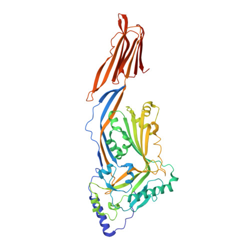
Crystal Structure of Listeriolysin O Reveals Molecular Details of Oligomerization and Pore Formation

Koester, S.Pee, K.V.Hudel, M.Leustik, M.Rhinow, D.Kuehlbrandt, W.Chakraborty, T.Yildiz, O.

(2014) Nat Commun 5: 3690

  • DOI: https://doi.org/10.1038/ncomms4690
  • Primary Citation of Related Structures:  
    4CDB

  • PubMed Abstract: 

    Listeriolysin O (LLO) is an essential virulence factor of Listeria monocytogenes that causes listeriosis. Listeria monocytogenes owes its ability to live within cells to the pH- and temperature-dependent pore-forming activity of LLO, which is unique among cholesterol-dependent cytolysins. LLO enables the bacteria to cross the phagosomal membrane and is also involved in activation of cellular processes, including the modulation of gene expression or intracellular Ca(2+) oscillations. Neither the pore-forming mechanism nor the mechanisms triggering the signalling processes in the host cell are known in detail. Here, we report the crystal structure of LLO, in which we identified regions important for oligomerization and pore formation. Mutants were characterized by determining their haemolytic and Ca(2+) uptake activity. We analysed the pore formation of LLO and its variants on erythrocyte ghosts by electron microscopy and show that pore formation requires precise interface interactions during toxin oligomerization on the membrane.


  • Organizational Affiliation

    Department of Structural Biology, Max Planck Institute of Biophysics, Max von Laue Str. 3, Frankfurt am Main 60438, Germany.


Macromolecules
Find similar proteins by:  (by identity cutoff)  |  3D Structure
Entity ID: 1
MoleculeChains Sequence LengthOrganismDetailsImage
LISTERIOLYSIN O488Listeria monocytogenesMutation(s): 0 
Membrane Entity: Yes 
UniProt
Find proteins for A0A0H3GD84 (Listeria monocytogenes serotype 1/2a (strain 10403S))
Explore A0A0H3GD84 
Go to UniProtKB:  A0A0H3GD84
Entity Groups  
Sequence Clusters30% Identity50% Identity70% Identity90% Identity95% Identity100% Identity
UniProt GroupA0A0H3GD84
Sequence Annotations
Expand
  • Reference Sequence
Small Molecules
Ligands 3 Unique
IDChains Name / Formula / InChI Key2D Diagram3D Interactions
TAM
Query on TAM

Download Ideal Coordinates CCD File 
U [auth A]TRIS(HYDROXYETHYL)AMINOMETHANE
C7 H17 N O3
GKODZWOPPOTFGA-UHFFFAOYSA-N
ACT
Query on ACT

Download Ideal Coordinates CCD File 
B [auth A],
C [auth A],
D [auth A],
E [auth A]
ACETATE ION
C2 H3 O2
QTBSBXVTEAMEQO-UHFFFAOYSA-M
NA
Query on NA

Download Ideal Coordinates CCD File 
F [auth A]
G [auth A]
H [auth A]
I [auth A]
J [auth A]
F [auth A],
G [auth A],
H [auth A],
I [auth A],
J [auth A],
K [auth A],
L [auth A],
M [auth A],
N [auth A],
O [auth A],
P [auth A],
Q [auth A],
R [auth A],
S [auth A],
T [auth A]
SODIUM ION
Na
FKNQFGJONOIPTF-UHFFFAOYSA-N
Experimental Data & Validation

Experimental Data

  • Method: X-RAY DIFFRACTION
  • Resolution: 2.15 Å
  • R-Value Free: 0.246 
  • R-Value Work: 0.194 
  • R-Value Observed: 0.196 
  • Space Group: P 21 21 21
Unit Cell:
Length ( Å )Angle ( ˚ )
a = 26.72α = 90
b = 85.15β = 90
c = 229.9γ = 90
Software Package:
Software NamePurpose
PHENIXrefinement
XDSdata reduction
XSCALEdata scaling
PHASERphasing

Structure Validation

View Full Validation Report


Entry History 

Deposition Data

Revision History  (Full details and data files)

  • Version 1.0: 2014-04-23
    Type: Initial release
  • Version 1.1: 2014-04-30
    Changes: Database references
  • Version 1.2: 2014-05-07
    Changes: Database references
  • Version 1.3: 2023-12-20
    Changes: Data collection, Database references, Derived calculations, Other, Refinement description