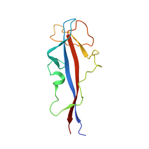
Crystal Structures of the Human Dysferlin Inner Dysf Domain
Sula, A., Cole, A.R., Yeats, C., Orengo, C., Keep, N.H.(2014) BMC Struct Biol 14: 3
- PubMed: 24438169 Search on PubMedSearch on PubMed Central
- DOI: https://doi.org/10.1186/1472-6807-14-3
- Primary Citation Related Structures:
4CAH, 4CAI - PubMed Abstract:
Mutations in dysferlin, the first protein linked with the cell membrane repair mechanism, causes a group of muscular dystrophies called dysferlinopathies. Dysferlin is a type two-anchored membrane protein, with a single C terminal trans-membrane helix, and most of the protein lying in cytoplasm. Dysferlin contains several C2 domains and two DysF domains which are nested one inside the other. Many pathogenic point mutations fall in the DysF domain region. We describe the crystal structure of the human dysferlin inner DysF domain with a resolution of 1.9 Ångstroms. Most of the pathogenic mutations are part of aromatic/arginine stacks that hold the domain in a folded conformation. The high resolution of the structure show that these interactions are a mixture of parallel ring/guanadinium stacking, perpendicular H bond stacking and aliphatic chain packing. The high resolution structure of the Dysferlin DysF domain gives a template on which to interpret in detail the pathogenic mutations that lead to disease.
- Crystallography, Biological Sciences, Institute for Structural and Molecular Biology, Birkbeck University of London, Malet Street, London WC1E 7HX, UK. n.keep@mail.cryst.bbk.ac.uk.
Organizational Affiliation: