Evidence that Gh115 Alpha-Glucuronidase Activity is Dependent on Conformational Flexibility
Rogowski, A., Basle, A., Farinas, C.S., Solovyova, A., Mortimer, J.C., Dupree, P., Gilbert, H.J., Bolam, D.N.(2014) J Biological Chem 289: 53
- PubMed: 24214982 Search on PubMedSearch on PubMed Central
- DOI: https://doi.org/10.1074/jbc.M113.525295
- Primary Citation Related Structures:
4C90, 4C91 - PubMed Abstract:
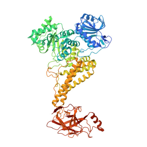
The microbial degradation of the plant cell wall is an important biological process that is highly relevant to environmentally significant industries such as the bioenergy and biorefining sectors. A major component of the wall is glucuronoxylan, a β1,4-linked xylose polysaccharide that is decorated with α-linked glucuronic and/or methylglucuronic acid (GlcA/MeGlcA). Recently three members of a glycoside hydrolase family, GH115, were shown to hydrolyze MeGlcA side chains from the internal regions of xylan, an activity that has not previously been described. Here we show that a dominant member of the human microbiota, Bacteroides ovatus, contains a GH115 enzyme, BoAgu115A, which displays glucuronoxylan α-(4-O-methyl)-glucuronidase activity. The enzyme is significantly more active against substrates in which the xylose decorated with GlcA/MeGlcA is flanked by one or more xylose residues. The crystal structure of BoAgu115A revealed a four-domain protein in which the active site, comprising a pocket that abuts a cleft-like structure, is housed in the second domain that adopts a TIM barrel-fold. The third domain, a five-helical bundle, and the C-terminal β-sandwich domain make inter-chain contacts leading to protein dimerization. Informed by the structure of the enzyme in complex with GlcA in its open ring form, in conjunction with mutagenesis studies, the potential substrate binding and catalytically significant amino acids were identified. Based on the catalytic importance of residues located on a highly flexible loop, the enzyme is required to undergo a substantial conformational change to form a productive Michaelis complex with glucuronoxylan.
- From the Institute for Cell and Molecular Biosciences, The Medical School, Newcastle University, Newcastle upon Tyne NE2 4HH United Kingdom and.
Organizational Affiliation: