4C6N | pdb_00004c6n

Crystal structure of the dihydroorotase domain of human CAD E1637T mutant bound to substrate at pH 6.0


Experimental Data Snapshot

  • Method: X-RAY DIFFRACTION
  • Resolution: 1.90 Å
  • R-Value Free: 
    0.170 (Depositor), 0.171 (DCC) 
  • R-Value Work: 
    0.146 (Depositor), 0.148 (DCC) 
  • R-Value Observed: 
    0.147 (Depositor) 

Starting Model: experimental
View more details

wwPDB Validation 3D Report Full Report

Validation slider image for 4C6N

Ligand Structure Quality Assessment 


This is version 1.2 of the entry. See complete history

Literature

Structure, Functional Characterization and Evolution of the Dihydroorotase Domain of Human Cad.

Grande-Garcia, A.Lallous, N.Diaz-Tejada, C.Ramon-Maiques, S.

(2014) Structure 22: 185

  • DOI: https://doi.org/10.1016/j.str.2013.10.016
  • Primary Citation Related Structures: 
    4BY3, 4C6B, 4C6C, 4C6D, 4C6E, 4C6F, 4C6I, 4C6J, 4C6K, 4C6L, 4C6M, 4C6N, 4C6O, 4C6P, 4C6Q

  • PubMed Abstract: 

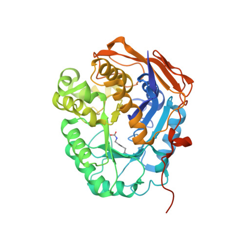
    Upregulation of CAD, the multifunctional protein that initiates and controls the de novo biosynthesis of pyrimidines in animals, is essential for cell proliferation. Deciphering the architecture and functioning of CAD is of interest for its potential usage as an antitumoral target. However, there is no detailed structural information about CAD other than that it self-assembles into hexamers of ∼1.5 MDa. Here we report the crystal structure and functional characterization of the dihydroorotase domain of human CAD. Contradicting all assumptions, the structure reveals an active site enclosed by a flexible loop with two Zn²⁺ ions bridged by a carboxylated lysine and a third Zn coordinating a rare histidinate ion. Site-directed mutagenesis and functional assays prove the involvement of the Zn and flexible loop in catalysis. Comparison with homologous bacterial enzymes supports a reclassification of the DHOase family and provides strong evidence against current models of the architecture of CAD.


  • Organizational Affiliation
    • Structural Bases of Genome Integrity Group, Structural Biology and Biocomputing Programme, Spanish National Cancer Research Centre (CNIO), 28029 Madrid, Spain.

Macromolecule Content 

  • Total Structure Weight: 43.33 kDa 
  • Atom Count: 3,213 
  • Modeled Residue Count: 363 
  • Deposited Residue Count: 393 
  • Unique protein chains: 1

Macromolecules

Find similar proteins by:|  3D Structure
Entity ID: 1
MoleculeChains  Sequence LengthOrganismDetailsImage
CAD PROTEIN393Homo sapiensMutation(s): 1 
EC: 3.5.2.3 (PDB Primary Data), 6.3.4.16 (UniProt), 6.3.5.5 (UniProt), 2.1.3.2 (UniProt), 3.5.1.2 (UniProt)
UniProt & NIH Common Fund Data Resources
Find proteins for P27708 (Homo sapiens)
Explore P27708 
Go to UniProtKB:  P27708
PHAROS:  P27708
GTEx:  ENSG00000084774 
Entity Groups
Sequence Clusters30% Identity50% Identity70% Identity90% Identity95% Identity100% Identity
UniProt GroupP27708
Sequence Annotations
Expand
Reference Sequence

Small Molecules

Modified Residues  1 Unique
IDChains TypeFormula2D DiagramParent
KCX
Query on KCX
A
L-PEPTIDE LINKINGC7 H14 N2 O4LYS

Experimental Data & Validation

Experimental Data

  • Method: X-RAY DIFFRACTION
  • Resolution: 1.90 Å
  • R-Value Free:  0.170 (Depositor), 0.171 (DCC) 
  • R-Value Work:  0.146 (Depositor), 0.148 (DCC) 
  • R-Value Observed: 0.147 (Depositor) 
Space Group: C 2 2 21
Unit Cell:
Length ( Å )Angle ( ˚ )
a = 82.1α = 90
b = 159.17β = 90
c = 60.97γ = 90
Software Package:
Software NamePurpose
PHENIXrefinement
XDSdata reduction
XSCALEdata scaling
PHASERphasing

Structure Validation

View Full Validation Report


Ligand Structure Quality Assessment 


Entry History 

Deposition Data

Revision History  (Full details and data files)

  • Version 1.0: 2014-02-05
    Type: Initial release
  • Version 1.1: 2014-02-19
    Changes: Database references
  • Version 1.2: 2023-12-20
    Changes: Data collection, Database references, Derived calculations, Other, Refinement description