4BKL

Crystal structure of the arthritogenic antibody M2139 (Fab fragment) in complex with the triple-helical J1 peptide


Experimental Data Snapshot

  • Method: X-RAY DIFFRACTION
  • Resolution: 3.25 Å
  • R-Value Free: 0.263 
  • R-Value Work: 0.222 
  • R-Value Observed: 0.224 

wwPDB Validation   3D Report Full Report


This is version 1.3 of the entry. See complete history


Literature

Epitope-Specific Antibody Response is Controlled by Immunoglobulin Vh Polymorphisms.

Raposo, B.Dobritzsch, D.Ge, C.Ekman, D.Xu, B.Lindh, I.Forster, M.Uysal, H.Nandakumar, K.S.Schneider, G.Holmdahl, R.

(2014) J Exp Med 211: 405

  • DOI: https://doi.org/10.1084/jem.20130968
  • Primary Citation of Related Structures:  
    4BKL

  • PubMed Abstract: 

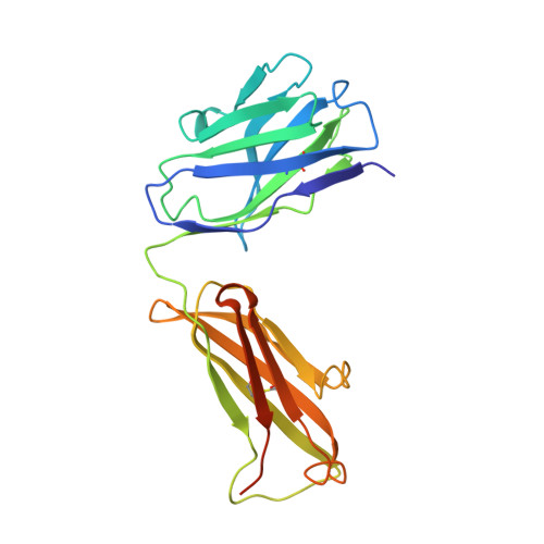
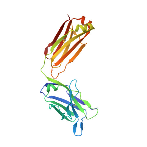
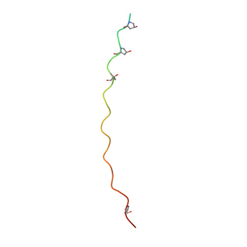
    Autoantibody formation is essential for the development of certain autoimmune diseases like rheumatoid arthritis (RA). Anti-type II collagen (CII) antibodies are found in RA patients; they interact with cartilage in vivo and are often highly pathogenic in the mouse. Autoreactivity to CII is directed to multiple epitopes and conserved between mice and humans. We have previously mapped the antibody response to CII in a heterogeneous stock cohort of mice, with a strong association with the IgH locus. We positioned the genetic polymorphisms and determined the structural requirements controlling antibody recognition of one of the major CII epitopes. Polymorphisms at positions S31R and W33T of the associated variable heavy chain (VH) allele were identified and confirmed by gene sequencing. The Fab fragment binding the J1 epitope was crystallized, and site-directed mutagenesis confirmed the importance of those two variants for antigen recognition. Back mutation to germline sequence provided evidence for a preexisting recognition of the J1 epitope. These data demonstrate a genetic association of epitope-specific antibody responses with specific VH alleles, and it highlights the importance of germline-encoded antibodies in the pathogenesis of antibody-mediated autoimmune diseases.


  • Organizational Affiliation

    Section for Medical Inflammation Research and 2 Section for Molecular Structural Biology, Department of Medical Biochemistry and Biophysics, Karolinska Institutet, 171 77 Stockholm, Sweden.


Macromolecules
Find similar proteins by:  (by identity cutoff)  |  3D Structure
Entity ID: 1
MoleculeChains Sequence LengthOrganismDetailsImage
M2139 FAB FRAGMENT HEAVY CHAIN231Mus musculusMutation(s): 0 
Entity Groups  
Sequence Clusters30% Identity50% Identity70% Identity90% Identity95% Identity100% Identity
Sequence Annotations
Expand
  • Reference Sequence
Find similar proteins by:  (by identity cutoff)  |  3D Structure
Entity ID: 2
MoleculeChains Sequence LengthOrganismDetailsImage
M2139 FAB FRAGMENT LIGHT CHAIN218Mus musculusMutation(s): 0 
Entity Groups  
Sequence Clusters30% Identity50% Identity70% Identity90% Identity95% Identity100% Identity
Sequence Annotations
Expand
  • Reference Sequence
Find similar proteins by:  (by identity cutoff)  |  3D Structure
Entity ID: 3
MoleculeChains Sequence LengthOrganismDetailsImage
J1 EPITOPEC [auth E],
D [auth F],
E [auth G]
37Mus musculusMutation(s): 0 
UniProt
Find proteins for P28481 (Mus musculus)
Explore P28481 
Go to UniProtKB:  P28481
Entity Groups  
Sequence Clusters30% Identity50% Identity70% Identity90% Identity95% Identity100% Identity
UniProt GroupP28481
Sequence Annotations
Expand
  • Reference Sequence
Small Molecules
Modified Residues  1 Unique
IDChains TypeFormula2D DiagramParent
HYP
Query on HYP
C [auth E],
D [auth F],
E [auth G]
L-PEPTIDE LINKINGC5 H9 N O3PRO
Experimental Data & Validation

Experimental Data

  • Method: X-RAY DIFFRACTION
  • Resolution: 3.25 Å
  • R-Value Free: 0.263 
  • R-Value Work: 0.222 
  • R-Value Observed: 0.224 
  • Space Group: P 31 2 1
Unit Cell:
Length ( Å )Angle ( ˚ )
a = 95.18α = 90
b = 95.18β = 90
c = 190.42γ = 120
Software Package:
Software NamePurpose
REFMACrefinement
MOSFLMdata reduction
SCALAdata scaling
PHASERphasing

Structure Validation

View Full Validation Report


Entry History 

Deposition Data

Revision History  (Full details and data files)

  • Version 1.0: 2014-02-05
    Type: Initial release
  • Version 1.1: 2014-03-05
    Changes: Database references
  • Version 1.2: 2014-03-26
    Changes: Database references
  • Version 1.3: 2023-12-20
    Changes: Data collection, Database references, Derived calculations, Other, Refinement description