4BJ0

Xyloglucan binding module (CBM4-2 X2-L110F) in complex with branched xyloses


Experimental Data Snapshot

  • Method: X-RAY DIFFRACTION
  • Resolution: 1.00 Å
  • R-Value Free: 0.129 
  • R-Value Work: 0.114 
  • R-Value Observed: 0.115 

wwPDB Validation   3D Report Full Report


Ligand Structure Quality Assessment 


This is version 2.1 of the entry. See complete history


Literature

Carbohydrate Binding Module Recognition of Xyloglucan Defined by Polar Contacts with Branching Xyloses and Ch-Pi Interactions.

von Schantz, L.Hakansson, M.Logan, D.T.Nordberg-Karlsson, E.Ohlin, M.

(2014) Proteins 82: 3466

  • DOI: https://doi.org/10.1002/prot.24700
  • Primary Citation of Related Structures:  
    4BJ0

  • PubMed Abstract: 

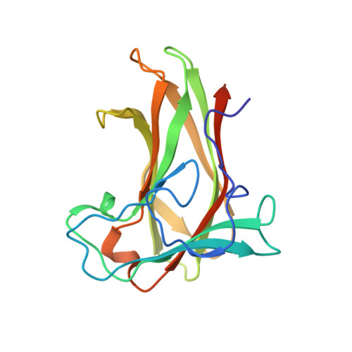
    Engineering of novel carbohydrate-binding proteins that can be utilized in various biochemical and biotechnical applications would benefit from a deeper understanding of the biochemical interactions that determine protein-carbohydrate specificity. In an effort to understand further the basis for specificity we present the crystal structure of the multi-specific carbohydrate-binding module (CBM) X-2 L110F bound to a branched oligomer of xyloglucan (XXXG). X-2 L110F is an engineered CBM that can recognize xyloglucan, xylans and β-glucans. The structural observations of the present study compared with previously reported structures of X-2 L110F in complex with linear oligomers, show that the π-surface of a phenylalanine, F110, allows for interactions with hydrogen atoms on both linear (xylopentaose and cellopentaose) and branched ligands (XXXG). Furthermore, X-2 L110F is shown to have a relatively flexible binding cleft, as illustrated in binding to XXXG. This branched ligand requires a set of reorientations of protein side chains Q72, N31, and R142, although these residues have previously been determined as important for binding to xylose oligomers by mediating polar contacts. The loss of these polar contacts is compensated for in binding to XXXG by polar interactions mediated by other protein residues, T74, R115, and Y149, which interact mainly with the branching xyloses of the xyloglucan oligomer. Taken together, the present study illustrates in structural detail how CH-π interactions can influence binding specificity and that flexibility is a key feature for the multi-specificity displayed by X-2 L110F, allowing for the accommodation of branched ligands.


  • Organizational Affiliation

    Department of Immunotechnology, Lund University, Medicon Village, SE-223 81 Lund, Sweden.


Macromolecules
Find similar proteins by:  (by identity cutoff)  |  3D Structure
Entity ID: 1
MoleculeChains Sequence LengthOrganismDetailsImage
XYLANASE176Rhodothermus marinusMutation(s): 7 
EC: 3.2.1.8
UniProt
Find proteins for Q6V8M0 (Rhodothermus marinus)
Explore Q6V8M0 
Go to UniProtKB:  Q6V8M0
Entity Groups  
Sequence Clusters30% Identity50% Identity70% Identity90% Identity95% Identity100% Identity
UniProt GroupQ6V8M0
Sequence Annotations
Expand
  • Reference Sequence
Oligosaccharides

Help

Entity ID: 2
MoleculeChains Length2D Diagram Glycosylation3D Interactions
alpha-D-xylopyranose-(1-6)-beta-D-glucopyranose-(1-4)-[alpha-D-xylopyranose-(1-6)]beta-D-glucopyranose-(1-4)-[alpha-D-xylopyranose-(1-6)]beta-D-glucopyranose-(1-4)-beta-D-glucopyranose
B
7N/A
Glycosylation Resources
GlyTouCan:  G24181CY
GlyCosmos:  G24181CY
Small Molecules
Experimental Data & Validation

Experimental Data

  • Method: X-RAY DIFFRACTION
  • Resolution: 1.00 Å
  • R-Value Free: 0.129 
  • R-Value Work: 0.114 
  • R-Value Observed: 0.115 
  • Space Group: P 21 21 2
Unit Cell:
Length ( Å )Angle ( ˚ )
a = 71.78α = 90
b = 48.79β = 90
c = 44.22γ = 90
Software Package:
Software NamePurpose
REFMACrefinement
XDSdata reduction
XSCALEdata scaling
PHASERphasing

Structure Validation

View Full Validation Report


Ligand Structure Quality Assessment 


Entry History 

Deposition Data

Revision History  (Full details and data files)

  • Version 1.0: 2014-04-23
    Type: Initial release
  • Version 1.1: 2014-10-29
    Changes: Database references
  • Version 1.2: 2014-12-03
    Changes: Database references
  • Version 1.3: 2018-01-17
    Changes: Data collection, Database references
  • Version 2.0: 2020-07-29
    Type: Remediation
    Reason: Carbohydrate remediation
    Changes: Atomic model, Data collection, Derived calculations, Other, Structure summary
  • Version 2.1: 2023-12-20
    Changes: Data collection, Database references, Refinement description, Structure summary