4BCP | pdb_00004bcp

Structure of CDK2 in complex with cyclin A and a 2-amino-4-heteroaryl- pyrimidine inhibitor


Experimental Data Snapshot

  • Method: X-RAY DIFFRACTION
  • Resolution: 2.26 Å
  • R-Value Free: 
    0.217 (Depositor), 0.204 (DCC) 
  • R-Value Work: 
    0.181 (Depositor) 
  • R-Value Observed: 
    0.183 (Depositor) 

wwPDB Validation 3D Report Full Report

Validation slider image for 4BCP

Ligand Structure Quality Assessment 


This is version 1.2 of the entry. See complete history

Literature

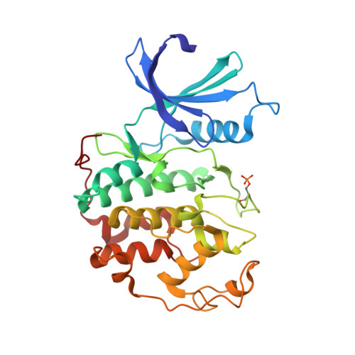
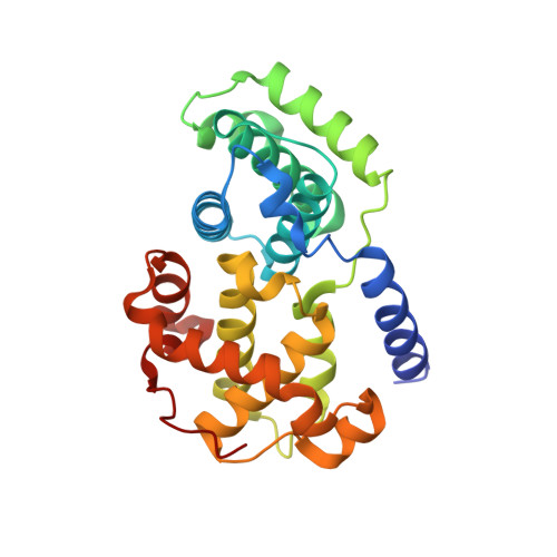
Substituted 4-(Thiazol-5-Yl)-2-(Phenylamino)Pyrimidines are Highly Active Cdk9 Inhibitors: Synthesis, X-Ray Crystal Structure, Sar and Anti-Cancer Activities.

Shao, H.Shi, S.Huang, S.Hole, A.Abbas, A.Y.Baumli, S.Liu, X.Lam, F.Foley, D.W.Fischer, P.M.Noble, M.Endicott, J.A.Pepper, C.Wang, S.

(2013) J Med Chem 56: 640

  • DOI: https://doi.org/10.1021/jm301475f
  • Primary Citation Related Structures: 
    4BCG, 4BCP

  • PubMed Abstract: 

    Cancer cells often have a high demand for antiapoptotic proteins in order to resist programmed cell death. CDK9 inhibition selectively targets survival proteins and reinstates apoptosis in cancer cells. We designed a series of 4-thiazol-2-anilinopyrimidine derivatives with functional groups attached to the C5-position of the pyrimidine or to the C4-thiazol moiety and investigated their effects on CDK9 potency and selectivity. One of the most selective compounds, 12u inhibits CDK9 with IC(50) = 7 nM and shows over 80-fold selectivity for CDK9 versus CDK2. X-ray crystal structures of 12u bound to CDK9 and CDK2 provide insights into the binding modes. This work, together with crystal structures of selected inhibitors in complex with both enzymes described in a companion paper, (34) provides a rationale for the observed SAR. 12u demonstrates potent anticancer activity against primary chronic lymphocytic leukemia cells with a therapeutic window 31- and 107-fold over those of normal B- and T-cells.


  • Organizational Affiliation
    • School of Pharmacy and Centre for Biomolecular Sciences, University of Nottingham, University Park, Nottingham NG7 2RD, UK.

Macromolecule Content 

  • Total Structure Weight: 129.87 kDa 
  • Atom Count: 9,233 
  • Modeled Residue Count: 1,081 
  • Deposited Residue Count: 1,124 
  • Unique protein chains: 2

Macromolecules

Find similar proteins by:|  3D Structure
Entity ID: 1
MoleculeChains  Sequence LengthOrganismDetailsImage
CYCLIN-DEPENDENT KINASE 2
A, C
300Homo sapiensMutation(s): 0 
EC: 2.7.11.22
UniProt & NIH Common Fund Data Resources
Find proteins for P24941 (Homo sapiens)
Explore P24941 
Go to UniProtKB:  P24941
PHAROS:  P24941
GTEx:  ENSG00000123374 
Entity Groups
Sequence Clusters30% Identity50% Identity70% Identity90% Identity95% Identity100% Identity
UniProt GroupP24941
Sequence Annotations
Expand
Reference Sequence
Find similar proteins by:|  3D Structure
Entity ID: 2
MoleculeChains  Sequence LengthOrganismDetailsImage
CYCLIN-A2
B, D
262Homo sapiensMutation(s): 0 
UniProt & NIH Common Fund Data Resources
Find proteins for P20248 (Homo sapiens)
Explore P20248 
Go to UniProtKB:  P20248
PHAROS:  P20248
GTEx:  ENSG00000145386 
Entity Groups
Sequence Clusters30% Identity50% Identity70% Identity90% Identity95% Identity100% Identity
UniProt GroupP20248
Sequence Annotations
Expand
Reference Sequence

Small Molecules

Ligands 3 Unique
IDChains Name / Formula / InChI Key2D Diagram3D Interactions
T3C

Query on T3C


Download:Ideal Coordinates CCD File
E [auth A],
I [auth C]
2-[[3-(1,4-diazepan-1-yl)phenyl]amino]-4-[4-methyl-2-(methylamino)-1,3-thiazol-5-yl]pyrimidine-5-carbonitrile
C21 H24 N8 S
BKOYUSKBPMSUBK-UHFFFAOYSA-N
SGM

Query on SGM


Download:Ideal Coordinates CCD File
G [auth B],
H [auth B],
K [auth D]
MONOTHIOGLYCEROL
C3 H8 O2 S
PJUIMOJAAPLTRJ-GSVOUGTGSA-N
SO4

Query on SO4


Download:Ideal Coordinates CCD File
F [auth B],
J [auth D]
SULFATE ION
O4 S
QAOWNCQODCNURD-UHFFFAOYSA-L
Modified Residues  1 Unique
IDChains TypeFormula2D DiagramParent
TPO
Query on TPO
A, C
L-PEPTIDE LINKINGC4 H10 N O6 PTHR

Experimental Data & Validation

Experimental Data

  • Method: X-RAY DIFFRACTION
  • Resolution: 2.26 Å
  • R-Value Free:  0.217 (Depositor), 0.204 (DCC) 
  • R-Value Work:  0.181 (Depositor) 
  • R-Value Observed: 0.183 (Depositor) 
Space Group: P 21 21 21
Unit Cell:
Length ( Å )Angle ( ˚ )
a = 73.81α = 90
b = 134.553β = 90
c = 149.168γ = 90
Software Package:
Software NamePurpose
PHENIXrefinement
PHASERphasing

Structure Validation

View Full Validation Report


Ligand Structure Quality Assessment 


Entry History 

Deposition Data

Revision History  (Full details and data files)

  • Version 1.0: 2013-04-17
    Type: Initial release
  • Version 1.1: 2013-08-07
    Changes: Refinement description
  • Version 1.2: 2024-11-13
    Changes: Data collection, Database references, Derived calculations, Other, Structure summary