Crystallization of Lysozyme with (R)-, (S)- and (Rs)-2-Methyl-2,4-Pentanediol
Stauber, M., Jakoncic, J., Berger, J., Karp, J.M., Axelbaum, A., Sastow, D., Buldyrev, S.V., Hrnjez, B.J., Asherie, N.(2015) Acta Crystallogr D Biol Crystallogr 71: 427
- PubMed: 25760593 Search on PubMedSearch on PubMed Central
- DOI: https://doi.org/10.1107/S1399004714025061
- Primary Citation Related Structures:
4B49, 4B4E, 4B4I, 4B4J - PubMed Abstract:
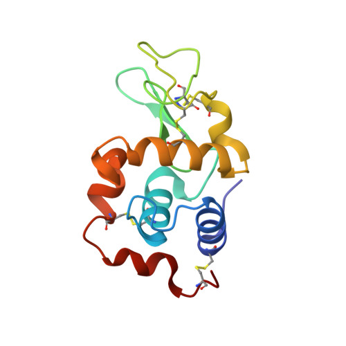
Chiral control of crystallization has ample precedent in the small-molecule world, but relatively little is known about the role of chirality in protein crystallization. In this study, lysozyme was crystallized in the presence of the chiral additive 2-methyl-2,4-pentanediol (MPD) separately using the R and S enantiomers as well as with a racemic RS mixture. Crystals grown with (R)-MPD had the most order and produced the highest resolution protein structures. This result is consistent with the observation that in the crystals grown with (R)-MPD and (RS)-MPD the crystal contacts are made by (R)-MPD, demonstrating that there is preferential interaction between lysozyme and this enantiomer. These findings suggest that chiral interactions are important in protein crystallization.
- Department of Physics, Yeshiva University, 2495 Amsterdam Avenue, New York, NY 10033-3312, USA.
Organizational Affiliation: