4AW8

X-ray structure of ZinT from Salmonella enterica in complex with zinc ion and PEG


Experimental Data Snapshot

  • Method: X-RAY DIFFRACTION
  • Resolution: 2.00 Å
  • R-Value Free: 0.257 
  • R-Value Work: 0.212 
  • R-Value Observed: 0.215 

wwPDB Validation   3D Report Full Report


This is version 1.3 of the entry. See complete history


Literature

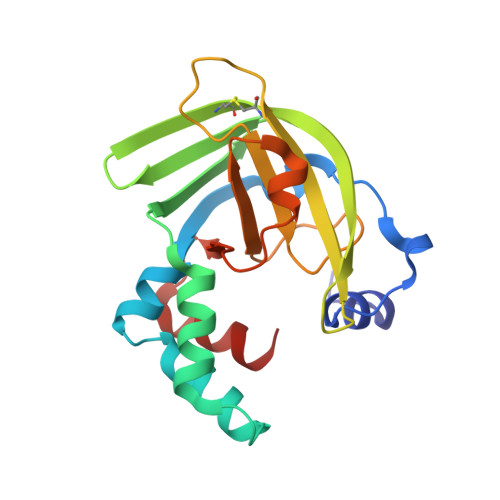
The Salmonella Enterica Zint Structure, Zinc Affinity and Interaction with the High-Affinity Uptake Protein Znua Provide Insight Into the Management of Periplasmic Zinc.

Ilari, A.Alaleona, F.Tria, G.Petrarca, P.Battistoni, A.Zamparelli, C.Verzili, D.Falconi, M.Chiancone, E.

(2014) Biochim Biophys Acta 1840: 535

  • DOI: https://doi.org/10.1016/j.bbagen.2013.10.010
  • Primary Citation of Related Structures:  
    4ARH, 4AW8, 4AYH

  • PubMed Abstract: 

    In Gram-negative bacteria the ZnuABC transporter ensures adequate zinc import in Zn(II)-poor environments, like those encountered by pathogens within the infected host. Recently, the metal-binding protein ZinT was suggested to operate as an accessory component of ZnuABC in periplasmic zinc recruitment. Since ZinT is known to form a ZinT-ZnuA complex in the presence of Zn(II) it was proposed to transfer Zn(II) to ZnuA. The present work was undertaken to test this claim. ZinT and its structural relationship with ZnuA have been characterized by multiple biophysical techniques (X-ray crystallography, SAXS, analytical ultracentrifugation, fluorescence spectroscopy). The metal-free and metal-bound crystal structures of Salmonella enterica ZinT show one Zn(II) binding site and limited structural changes upon metal removal. Spectroscopic titrations with Zn(II) yield a KD value of 22±2nM for ZinT, while those with ZnuA point to one high affinity (KD<20nM) and one low affinity Zn(II) binding site (KD in the micromolar range). Sedimentation velocity experiments established that Zn(II)-bound ZinT interacts with ZnuA, whereas apo-ZinT does not. The model of the ZinT-ZnuA complex derived from small angle X-ray scattering experiments points to a disposition that favors metal transfer as the metal binding cavities of the two proteins face each other. ZinT acts as a Zn(II)-buffering protein that delivers Zn(II) to ZnuA. Knowledge of the ZinT-ZnuA relationship is crucial for understanding bacterial Zn(II) uptake.


  • Organizational Affiliation

    CNR Institute of Molecular Biology and Pathology and Department of Biochemical Sciences, "Sapienza" University of Rome, Piazzale A. Moro 5, 00185 Rome, Italy.


Macromolecules
Find similar proteins by:  (by identity cutoff)  |  3D Structure
Entity ID: 1
MoleculeChains Sequence LengthOrganismDetailsImage
Metal-binding protein ZinT186Salmonella enterica subsp. enterica serovar 4,[5],12:i:- str. CVM23701Mutation(s): 0 
Gene Names: zinTDBP43_03300DVH39_02025DVH40_02355DVH41_01240DVH48_05500
UniProt
Find proteins for A0A315GY36 (Salmonella enterica subsp. enterica serovar 4,[5],12:i:-)
Explore A0A315GY36 
Go to UniProtKB:  A0A315GY36
Entity Groups  
Sequence Clusters30% Identity50% Identity70% Identity90% Identity95% Identity100% Identity
UniProt GroupA0A315GY36
Sequence Annotations
Expand
  • Reference Sequence
Small Molecules
Ligands 4 Unique
IDChains Name / Formula / InChI Key2D Diagram3D Interactions
PG6
Query on PG6

Download Ideal Coordinates CCD File 
H [auth A]1-(2-METHOXY-ETHOXY)-2-{2-[2-(2-METHOXY-ETHOXY]-ETHOXY}-ETHANE
C12 H26 O6
DMDPGPKXQDIQQG-UHFFFAOYSA-N
SO4
Query on SO4

Download Ideal Coordinates CCD File 
C [auth A],
D [auth A],
E [auth A],
F [auth A],
G [auth A]
SULFATE ION
O4 S
QAOWNCQODCNURD-UHFFFAOYSA-L
ZN
Query on ZN

Download Ideal Coordinates CCD File 
B [auth A]ZINC ION
Zn
PTFCDOFLOPIGGS-UHFFFAOYSA-N
NA
Query on NA

Download Ideal Coordinates CCD File 
I [auth A],
J [auth A]
SODIUM ION
Na
FKNQFGJONOIPTF-UHFFFAOYSA-N
Binding Affinity Annotations 
IDSourceBinding Affinity
ZN PDBBind:  4AW8 Kd: 22 (nM) from 1 assay(s)
Experimental Data & Validation

Experimental Data

  • Method: X-RAY DIFFRACTION
  • Resolution: 2.00 Å
  • R-Value Free: 0.257 
  • R-Value Work: 0.212 
  • R-Value Observed: 0.215 
  • Space Group: P 61 2 2
Unit Cell:
Length ( Å )Angle ( ˚ )
a = 58.54α = 90
b = 58.54β = 90
c = 290.37γ = 120
Software Package:
Software NamePurpose
REFMACrefinement
MOSFLMdata reduction
SCALAdata scaling
MOLREPphasing

Structure Validation

View Full Validation Report


Entry History 

Deposition Data

Revision History  (Full details and data files)

  • Version 1.0: 2013-06-19
    Type: Initial release
  • Version 1.1: 2013-12-25
    Changes: Database references
  • Version 1.2: 2019-10-16
    Changes: Data collection, Database references, Other, Source and taxonomy, Structure summary
  • Version 1.3: 2024-05-01
    Changes: Data collection, Database references, Derived calculations, Refinement description