4AVC

Crystal structure of protein lysine acetyltransferase Rv0998 in complex with acetyl CoA and cAMP


Experimental Data Snapshot

  • Method: X-RAY DIFFRACTION
  • Resolution: 2.81 Å
  • R-Value Free: 0.267 
  • R-Value Work: 0.199 
  • R-Value Observed: 0.202 

wwPDB Validation   3D Report Full Report


This is version 1.1 of the entry. See complete history


Literature

Cyclic AMP Regulation of Protein Lysine Acetylation in Mycobacterium Tuberculosis.

Lee, H.J.Lang, P.T.Fortune, S.M.Sassetti, C.M.Alber, T.

(2012) Nat Struct Mol Biol 19: 811

  • DOI: https://doi.org/10.1038/nsmb.2318
  • Primary Citation of Related Structures:  
    4AVA, 4AVB, 4AVC

  • PubMed Abstract: 

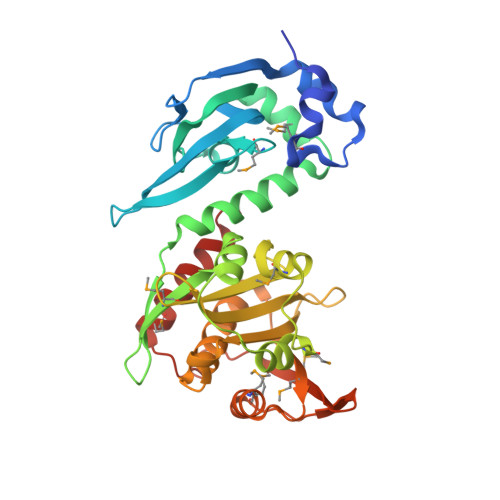
    Protein lysine acetylation networks can regulate central processes such as carbon metabolism and gene expression in bacteria. In Escherichia coli, cyclic AMP (cAMP) regulates protein lysine acetyltransferase (PAT) activity at the transcriptional level, but in Mycobacterium tuberculosis, fusion of a cyclic nucleotide-binding domain to a Gcn5-like PAT domain enables direct cAMP control of protein acetylation. Here we describe the allosteric activation mechanism of M. tuberculosis PAT. The crystal structures of the autoinhibited and cAMP-activated PAT reveal that cAMP binds to a cryptic site in the regulatory domain that is over 32 Å from the catalytic site. An extensive conformational rearrangement relieves this autoinhibition by means of a substrate-mimicking lid that covers the protein-substrate binding surface. A steric double latch couples the domains by harnessing a classic, cAMP-mediated conformational switch. The structures suggest general features that enable the evolution of long-range communication between linked domains.


  • Organizational Affiliation

    Department of Molecular and Cell Biology, University of California, Berkeley, California, USA.


Macromolecules
Find similar proteins by:  (by identity cutoff)  |  3D Structure
Entity ID: 1
MoleculeChains Sequence LengthOrganismDetailsImage
LYSINE ACETYLTRANSFERASE
A, B
333Mycobacterium tuberculosis H37RvMutation(s): 0 
UniProt
Find proteins for O05581 (Mycobacterium tuberculosis (strain ATCC 25618 / H37Rv))
Explore O05581 
Go to UniProtKB:  O05581
Entity Groups  
Sequence Clusters30% Identity50% Identity70% Identity90% Identity95% Identity100% Identity
UniProt GroupO05581
Sequence Annotations
Expand
  • Reference Sequence
Small Molecules
Ligands 4 Unique
IDChains Name / Formula / InChI Key2D Diagram3D Interactions
ACO
Query on ACO

Download Ideal Coordinates CCD File 
D [auth A],
G [auth B]
ACETYL COENZYME *A
C23 H38 N7 O17 P3 S
ZSLZBFCDCINBPY-ZSJPKINUSA-N
CMP
Query on CMP

Download Ideal Coordinates CCD File 
C [auth A],
F [auth B]
ADENOSINE-3',5'-CYCLIC-MONOPHOSPHATE
C10 H12 N5 O6 P
IVOMOUWHDPKRLL-KQYNXXCUSA-N
PEG
Query on PEG

Download Ideal Coordinates CCD File 
E [auth A],
H [auth B],
I [auth B]
DI(HYDROXYETHYL)ETHER
C4 H10 O3
MTHSVFCYNBDYFN-UHFFFAOYSA-N
EDO
Query on EDO

Download Ideal Coordinates CCD File 
J [auth B]1,2-ETHANEDIOL
C2 H6 O2
LYCAIKOWRPUZTN-UHFFFAOYSA-N
Modified Residues  1 Unique
IDChains TypeFormula2D DiagramParent
MSE
Query on MSE
A, B
L-PEPTIDE LINKINGC5 H11 N O2 SeMET
Experimental Data & Validation

Experimental Data

  • Method: X-RAY DIFFRACTION
  • Resolution: 2.81 Å
  • R-Value Free: 0.267 
  • R-Value Work: 0.199 
  • R-Value Observed: 0.202 
  • Space Group: P 1 21 1
Unit Cell:
Length ( Å )Angle ( ˚ )
a = 68.661α = 90
b = 50.151β = 106.63
c = 110.102γ = 90
Software Package:
Software NamePurpose
PHENIXrefinement
HKL-2000data reduction
SCALEPACKdata scaling
PHENIXphasing

Structure Validation

View Full Validation Report


Entry History 

Deposition Data

Revision History  (Full details and data files)

  • Version 1.0: 2012-07-11
    Type: Initial release
  • Version 1.1: 2012-08-22
    Changes: Database references