4AU9 | pdb_00004au9

Crystal Structure of a Fungal DyP-Type Peroxidase from Auricularia auricula-judae


Experimental Data Snapshot

  • Method: X-RAY DIFFRACTION
  • Resolution: 2.10 Å
  • R-Value Free: 
    0.261 (Depositor), 0.261 (DCC) 
  • R-Value Work: 
    0.180 (Depositor), 0.181 (DCC) 
  • R-Value Observed: 
    0.184 (Depositor) 

Starting Model: experimental
View more details

wwPDB Validation 3D Report Full Report

Validation slider image for 4AU9

Ligand Structure Quality Assessment 


This is version 2.2 of the entry. See complete history

Literature

First Crystal Structure of a Fungal High-Redox Potential Dye-Decolorizing Peroxidase: Substrate Interaction Sites and Long-Range Electron Transfer.

Strittmatter, E.Liers, C.Ullrich, R.Wachter, S.Hofrichter, M.Plattner, D.A.Piontek, K.

(2013) J Biological Chem 288: 4095

  • DOI: https://doi.org/10.1074/jbc.M112.400176
  • Primary Citation Related Structures: 
    4AU9

  • PubMed Abstract: 

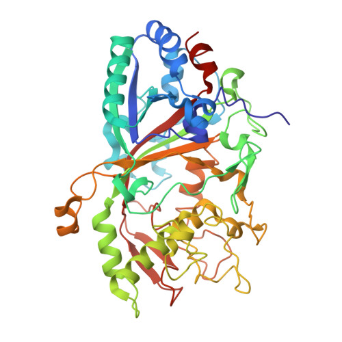
    Dye-decolorizing peroxidases (DyPs) belong to the large group of heme peroxidases. They utilize hydrogen peroxide to catalyze oxidations of various organic compounds. AauDyPI from Auricularia auricula-judae (fungi) was crystallized, and its crystal structure was determined at 2.1 Å resolution. The mostly helical structure also shows a β-sheet motif typical for DyPs and Cld (chlorite dismutase)-related structures and includes the complete polypeptide chain. At the distal side of the heme molecule, a flexible aspartate residue (Asp-168) plays a key role in catalysis. It guides incoming hydrogen peroxide toward the heme iron and mediates proton rearrangement in the process of Compound I formation. Afterward, its side chain changes its conformation, now pointing toward the protein backbone. We propose an extended functionality of Asp-168, which acts like a gatekeeper by altering the width of the heme cavity access channel. Chemical modifications of potentially redox-active amino acids show that a tyrosine is involved in substrate interaction. Using spin-trapping experiments, a transient radical on the surface-exposed Tyr-337 was identified as the oxidation site for bulky substrates. A possible long-range electron transfer pathway from the surface of the enzyme to the redox cofactor (heme) is discussed.


  • Organizational Affiliation
    • Institute of Organic Chemistry, University of Freiburg, Albertstrasse 21, 79104 Freiburg, Germany.

Macromolecule Content 

  • Total Structure Weight: 97.66 kDa 
  • Atom Count: 7,571 
  • Modeled Residue Count: 896 
  • Deposited Residue Count: 896 
  • Unique protein chains: 1

Macromolecules

Find similar proteins by:|  3D Structure
Entity ID: 1
MoleculeChains  Sequence LengthOrganismDetailsImage
DYP-TYPE PEROXIDASE I
A, B
448Auricularia auricula-judaeMutation(s): 0 
EC: 1.11.1.19 (PDB Primary Data), 1.11.1.7 (UniProt)
UniProt
Find proteins for I2DBY1 (Auricularia auricula-judae)
Explore I2DBY1 
Go to UniProtKB:  I2DBY1
Entity Groups
Sequence Clusters30% Identity50% Identity70% Identity90% Identity95% Identity100% Identity
UniProt GroupI2DBY1
Glycosylation
Glycosylation Sites: 2
Sequence Annotations
Expand
Reference Sequence

Oligosaccharides

Help  
Entity ID: 2
MoleculeChains Length2D Diagram GlycosylationD Interactions
beta-D-mannopyranose-(1-4)-2-acetamido-2-deoxy-beta-D-glucopyranose-(1-4)-2-acetamido-2-deoxy-beta-D-glucopyranose
C
3N-Glycosylation
Glycosylation Resources
GlyTouCan: G15407YE
GlyCosmos: G15407YE
GlyGen: G15407YE
Entity ID: 3
MoleculeChains Length2D Diagram GlycosylationD Interactions
2-acetamido-2-deoxy-beta-D-glucopyranose-(1-4)-2-acetamido-2-deoxy-beta-D-glucopyranose
D
2N-Glycosylation
Glycosylation Resources
GlyTouCan: G42666HT
GlyCosmos: G42666HT
GlyGen: G42666HT

Small Molecules

Ligands 5 Unique
IDChains Name / Formula / InChI Key2D Diagram3D Interactions
HEM

Query on HEM


Download:Ideal Coordinates CCD File
E [auth A],
L [auth B]
PROTOPORPHYRIN IX CONTAINING FE
C34 H32 Fe N4 O4
KABFMIBPWCXCRK-RGGAHWMASA-L
NAG

Query on NAG


Download:Ideal Coordinates CCD File
F [auth A],
G [auth A],
M [auth B]
2-acetamido-2-deoxy-beta-D-glucopyranose
C8 H15 N O6
OVRNDRQMDRJTHS-FMDGEEDCSA-N
TAM

Query on TAM


Download:Ideal Coordinates CCD File
O [auth B]TRIS(HYDROXYETHYL)AMINOMETHANE
C7 H17 N O3
GKODZWOPPOTFGA-UHFFFAOYSA-N
GOL

Query on GOL


Download:Ideal Coordinates CCD File
H [auth A]
I [auth A]
J [auth A]
N [auth B]
P [auth B]
H [auth A],
I [auth A],
J [auth A],
N [auth B],
P [auth B],
Q [auth B]
GLYCEROL
C3 H8 O3
PEDCQBHIVMGVHV-UHFFFAOYSA-N
ACT

Query on ACT


Download:Ideal Coordinates CCD File
K [auth A]ACETATE ION
C2 H3 O2
QTBSBXVTEAMEQO-UHFFFAOYSA-M

Experimental Data & Validation

Experimental Data

  • Method: X-RAY DIFFRACTION
  • Resolution: 2.10 Å
  • R-Value Free:  0.261 (Depositor), 0.261 (DCC) 
  • R-Value Work:  0.180 (Depositor), 0.181 (DCC) 
  • R-Value Observed: 0.184 (Depositor) 
Space Group: P 1 21 1
Unit Cell:
Length ( Å )Angle ( ˚ )
a = 66.599α = 90
b = 46.689β = 91.35
c = 141.198γ = 90
Software Package:
Software NamePurpose
REFMACrefinement
XDSdata reduction
XDSdata scaling
PHASERphasing

Structure Validation

View Full Validation Report


Ligand Structure Quality Assessment 


Entry History 

Deposition Data

Revision History  (Full details and data files)

  • Version 1.0: 2012-12-26
    Type: Initial release
  • Version 1.1: 2013-02-20
    Changes: Database references
  • Version 1.2: 2015-01-14
    Changes: Atomic model, Derived calculations
  • Version 2.0: 2020-07-29
    Type: Remediation
    Reason: Carbohydrate remediation
    Changes: Advisory, Atomic model, Data collection, Derived calculations, Other, Structure summary
  • Version 2.1: 2023-12-20
    Changes: Data collection, Database references, Refinement description, Structure summary
  • Version 2.2: 2024-11-13
    Changes: Structure summary