4AHV

Parallel screening of a low molecular weight compound library: do differences in methodology affect hit identification


Experimental Data Snapshot

  • Method: X-RAY DIFFRACTION
  • Resolution: 1.80 Å
  • R-Value Free: 0.224 
  • R-Value Work: 0.175 
  • R-Value Observed: 0.177 

wwPDB Validation   3D Report Full Report


Ligand Structure Quality Assessment 


This is version 1.4 of the entry. See complete history


Literature

Parallel Screening of Low Molecular Weight Fragment Libraries: Do Differences in Methodology Affect Hit Identification?

Wielens, J.Headey, S.J.Rhodes, D.I.Mulder, R.J.Dolezal, O.Deadman, J.J.Newman, J.Chalmers, D.K.Parker, M.W.Peat, T.S.Scanlon, M.J.

(2013) J Biomol Screen 18: 147

  • DOI: https://doi.org/10.1177/1087057112465979
  • Primary Citation of Related Structures:  
    3VQ4, 3VQ5, 3VQ6, 3VQ7, 3VQ8, 3VQ9, 3VQA, 3VQB, 3VQC, 3VQD, 3VQE, 3VQP, 3VQQ, 4AH9, 4AHR, 4AHS, 4AHT, 4AHU, 4AHV

  • PubMed Abstract: 

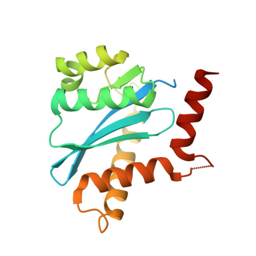
    Fragment screening is becoming widely accepted as a technique to identify hit compounds for the development of novel lead compounds. In neighboring laboratories, we have recently, and independently, performed a fragment screening campaign on the HIV-1 integrase core domain (IN) using similar commercially purchased fragment libraries. The two campaigns used different screening methods for the preliminary identification of fragment hits; one used saturation transfer difference nuclear magnetic resonance spectroscopy (STD-NMR), and the other used surface plasmon resonance (SPR) spectroscopy. Both initial screens were followed by X-ray crystallography. Using the STD-NMR/X-ray approach, 15 IN/fragment complexes were identified, whereas the SPR/X-ray approach found 6 complexes. In this article, we compare the approaches that were taken by each group and the results obtained, and we look at what factors could potentially influence the final results. We find that despite using different approaches with little overlap of initial hits, both approaches identified binding sites on IN that provided a basis for fragment-based lead discovery and further lead development. Comparison of hits identified in the two studies highlights a key role for both the conditions under which fragment binding is measured and the criteria selected to classify hits.


  • Organizational Affiliation

    St. Vincent's Institute, Fitzroy, Victoria, Australia.


Macromolecules
Find similar proteins by:  (by identity cutoff)  |  3D Structure
Entity ID: 1
MoleculeChains Sequence LengthOrganismDetailsImage
INTEGRASE
A, B
183Human immunodeficiency virusMutation(s): 3 
EC: 2.7.7.1
UniProt
Find proteins for P12497 (Human immunodeficiency virus type 1 group M subtype B (isolate NY5))
Explore P12497 
Go to UniProtKB:  P12497
Entity Groups  
Sequence Clusters30% Identity50% Identity70% Identity90% Identity95% Identity100% Identity
UniProt GroupP12497
Sequence Annotations
Expand
  • Reference Sequence
Small Molecules
Ligands 5 Unique
IDChains Name / Formula / InChI Key2D Diagram3D Interactions
Z5P
Query on Z5P

Download Ideal Coordinates CCD File 
L [auth A],
S [auth B]
1-[2-(1H-pyrazol-1-yl)phenyl]methanamine
C10 H11 N3
SJMJUZRSTJBVPG-UHFFFAOYSA-N
TAM
Query on TAM

Download Ideal Coordinates CCD File 
K [auth A],
R [auth B]
TRIS(HYDROXYETHYL)AMINOMETHANE
C7 H17 N O3
GKODZWOPPOTFGA-UHFFFAOYSA-N
SO4
Query on SO4

Download Ideal Coordinates CCD File 
C [auth A]
D [auth A]
E [auth A]
M [auth B]
N [auth B]
C [auth A],
D [auth A],
E [auth A],
M [auth B],
N [auth B],
O [auth B]
SULFATE ION
O4 S
QAOWNCQODCNURD-UHFFFAOYSA-L
EDO
Query on EDO

Download Ideal Coordinates CCD File 
H [auth A],
I [auth A],
J [auth A],
P [auth B],
Q [auth B]
1,2-ETHANEDIOL
C2 H6 O2
LYCAIKOWRPUZTN-UHFFFAOYSA-N
ACY
Query on ACY

Download Ideal Coordinates CCD File 
F [auth A],
G [auth A]
ACETIC ACID
C2 H4 O2
QTBSBXVTEAMEQO-UHFFFAOYSA-N
Binding Affinity Annotations 
IDSourceBinding Affinity
Z5P PDBBind:  4AHV Kd: 1.50e+7 (nM) from 1 assay(s)
Experimental Data & Validation

Experimental Data

  • Method: X-RAY DIFFRACTION
  • Resolution: 1.80 Å
  • R-Value Free: 0.224 
  • R-Value Work: 0.175 
  • R-Value Observed: 0.177 
  • Space Group: P 31
Unit Cell:
Length ( Å )Angle ( ˚ )
a = 70.923α = 90
b = 70.923β = 90
c = 66.69γ = 120
Software Package:
Software NamePurpose
REFMACrefinement
MOSFLMdata reduction
SCALAdata scaling
PHASERphasing

Structure Validation

View Full Validation Report


Ligand Structure Quality Assessment 


Entry History 

Deposition Data

Revision History  (Full details and data files)

  • Version 1.0: 2012-12-19
    Type: Initial release
  • Version 1.1: 2013-01-30
    Changes: Database references
  • Version 1.2: 2019-05-08
    Changes: Data collection, Experimental preparation, Other
  • Version 1.3: 2019-05-15
    Changes: Data collection, Experimental preparation
  • Version 1.4: 2023-12-20
    Changes: Data collection, Database references, Derived calculations, Other, Refinement description