4AAS | pdb_00004aas

ATP-triggered molecular mechanics of the chaperonin GroEL


Experimental Data Snapshot

  • Method: ELECTRON MICROSCOPY
  • Resolution: 8.50 Å
  • Aggregation State: PARTICLE 
  • Reconstruction Method: SINGLE PARTICLE 

Starting Model: experimental
View more details

wwPDB Validation 3D Report Full Report

Validation slider image for 4AAS

This is version 1.3 of the entry. See complete history

Literature

ATP-Triggered Conformational Changes Delineate Substrate-Binding and -Folding Mechanics of the Groel Chaperonin.

Clare, D.K.Vasishtan, D.Stagg, S.Quispe, J.Farr, G.W.Topf, M.Horwich, A.L.Saibil, H.R.

(2012) Cell 149: 113

  • DOI: https://doi.org/10.1016/j.cell.2012.02.047
  • Primary Citation Related Structures: 
    4AAQ, 4AAR, 4AAS, 4AAU, 4AB2, 4AB3

  • PubMed Abstract: 

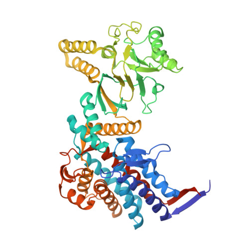
    The chaperonin GroEL assists the folding of nascent or stress-denatured polypeptides by actions of binding and encapsulation. ATP binding initiates a series of conformational changes triggering the association of the cochaperonin GroES, followed by further large movements that eject the substrate polypeptide from hydrophobic binding sites into a GroES-capped, hydrophilic folding chamber. We used cryo-electron microscopy, statistical analysis, and flexible fitting to resolve a set of distinct GroEL-ATP conformations that can be ordered into a trajectory of domain rotation and elevation. The initial conformations are likely to be the ones that capture polypeptide substrate. Then the binding domains extend radially to separate from each other but maintain their binding surfaces facing the cavity, potentially exerting mechanical force upon kinetically trapped, misfolded substrates. The extended conformation also provides a potential docking site for GroES, to trigger the final, 100° domain rotation constituting the "power stroke" that ejects substrate into the folding chamber.


  • Organizational Affiliation
    • Crystallography and Institute of Structural and Molecular Biology, Birkbeck College, University of London, Malet Street, London WC1E 7HX, UK.

Macromolecule Content 

  • Total Structure Weight: 807.25 kDa 
  • Atom Count: 54,062 
  • Modeled Residue Count: 7,336 
  • Deposited Residue Count: 7,672 
  • Unique protein chains: 1

Macromolecules

Find similar proteins by:|  3D Structure
Entity ID: 1
MoleculeChains  Sequence LengthOrganismDetailsImage
60 KDA CHAPERONIN
A, B, C, D, E
A, B, C, D, E, F, G, H, I, J, K, L, M, N
548Escherichia coliMutation(s): 1 
EC: 5.6.1.7
UniProt
Find proteins for P0A6F5 (Escherichia coli (strain K12))
Explore P0A6F5 
Go to UniProtKB:  P0A6F5
Entity Groups
Sequence Clusters30% Identity50% Identity70% Identity90% Identity95% Identity100% Identity
UniProt GroupP0A6F5
Sequence Annotations
Expand
Reference Sequence

Small Molecules

Ligands 3 Unique
IDChains Name / Formula / InChI Key2D Diagram3D Interactions
ATP

Query on ATP


Download:Ideal Coordinates CCD File
CA [auth E]
FA [auth F]
IA [auth G]
Q [auth A]
T [auth B]
CA [auth E],
FA [auth F],
IA [auth G],
Q [auth A],
T [auth B],
W [auth C],
Z [auth D]
ADENOSINE-5'-TRIPHOSPHATE
C10 H16 N5 O13 P3
ZKHQWZAMYRWXGA-KQYNXXCUSA-N
PO4

Query on PO4


Download:Ideal Coordinates CCD File
AA [auth E]
DA [auth F]
GA [auth G]
O [auth A]
R [auth B]
AA [auth E],
DA [auth F],
GA [auth G],
O [auth A],
R [auth B],
U [auth C],
X [auth D]
PHOSPHATE ION
O4 P
NBIIXXVUZAFLBC-UHFFFAOYSA-K
MG

Query on MG


Download:Ideal Coordinates CCD File
BA [auth E]
EA [auth F]
HA [auth G]
P [auth A]
S [auth B]
BA [auth E],
EA [auth F],
HA [auth G],
P [auth A],
S [auth B],
V [auth C],
Y [auth D]
MAGNESIUM ION
Mg
JLVVSXFLKOJNIY-UHFFFAOYSA-N

Experimental Data & Validation

Experimental Data

  • Method: ELECTRON MICROSCOPY
  • Resolution: 8.50 Å
  • Aggregation State: PARTICLE 
  • Reconstruction Method: SINGLE PARTICLE 
EM Software:
TaskSoftware PackageVersion
RECONSTRUCTIONIMAGIC
RECONSTRUCTIONSPIDER

Structure Validation

View Full Validation Report


Entry History 

Deposition Data

Revision History  (Full details and data files)

  • Version 1.0: 2012-12-12
    Type: Initial release
  • Version 1.1: 2013-09-25
    Changes: Other, Structure summary
  • Version 1.2: 2017-08-23
    Changes: Data collection, Refinement description
  • Version 1.3: 2024-05-08
    Changes: Data collection, Database references, Derived calculations, Refinement description