4A3P

Structure of USP15 DUSP-UBL deletion mutant


Experimental Data Snapshot

  • Method: X-RAY DIFFRACTION
  • Resolution: 1.40 Å
  • R-Value Free: 0.221 
  • R-Value Work: 0.153 
  • R-Value Observed: 0.157 

wwPDB Validation   3D Report Full Report


This is version 1.1 of the entry. See complete history


Literature

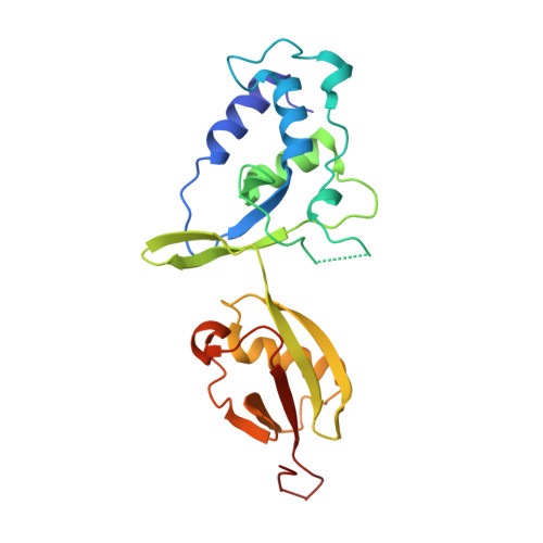
Structural Variability of the Ubiquitin Specific Protease Dusp-Ubl Double Domains.

Elliott, P.R.Liu, H.Pastok, M.W.Grossmann, G.J.Rigden, D.J.Clague, M.J.Urbe, S.Barsukov, I.L.

(2011) FEBS Lett 585: 3385

  • DOI: https://doi.org/10.1016/j.febslet.2011.09.040
  • Primary Citation of Related Structures:  
    3PV1, 4A3O, 4A3P

  • PubMed Abstract: 

    USP4, 11 and 15 are three closely related paralogues of the ubiquitin specific protease (USP) family of deubiquitinating enzymes. The DUSP domain and the UBL domain in these proteins are juxtaposed which may provide a functional unit conferring specificity. We determined the structures of the USP15 DUSP-UBL double domain unit in monomeric and dimeric states. We then conducted comparative analysis of the structural and physical properties of all three DUSP-UBL units. We identified structural features that dictate different dispositions between constituent domains, which in turn may influence respective binding properties.


  • Organizational Affiliation

    The University of Liverpool, Institute of Integrative Biology, Biosciences Building, Crown Street, Liverpool L69 7ZB, United Kingdom. P.R.Elliott@liv.ac.uk


Macromolecules
Find similar proteins by:  (by identity cutoff)  |  3D Structure
Entity ID: 1
MoleculeChains Sequence LengthOrganismDetailsImage
UBIQUITIN CARBOXYL-TERMINAL HYDROLASE 15217Homo sapiensMutation(s): 0 
EC: 3.4.19.12
UniProt & NIH Common Fund Data Resources
Find proteins for Q9Y4E8 (Homo sapiens)
Explore Q9Y4E8 
Go to UniProtKB:  Q9Y4E8
PHAROS:  Q9Y4E8
GTEx:  ENSG00000135655 
Entity Groups  
Sequence Clusters30% Identity50% Identity70% Identity90% Identity95% Identity100% Identity
UniProt GroupQ9Y4E8
Sequence Annotations
Expand
  • Reference Sequence
Experimental Data & Validation

Experimental Data

  • Method: X-RAY DIFFRACTION
  • Resolution: 1.40 Å
  • R-Value Free: 0.221 
  • R-Value Work: 0.153 
  • R-Value Observed: 0.157 
  • Space Group: P 1 21 1
Unit Cell:
Length ( Å )Angle ( ˚ )
a = 44.95α = 90
b = 44.25β = 104.33
c = 56.15γ = 90
Software Package:
Software NamePurpose
REFMACrefinement
MOSFLMdata reduction
SCALAdata scaling
PHASERphasing

Structure Validation

View Full Validation Report


Entry History 

Deposition Data

Revision History  (Full details and data files)

  • Version 1.0: 2011-11-16
    Type: Initial release
  • Version 1.1: 2023-12-20
    Changes: Data collection, Database references, Derived calculations, Other, Refinement description