4A0J

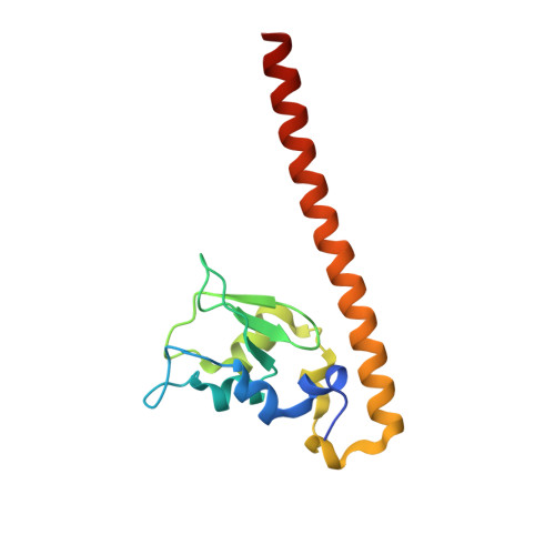
Crystal structure of Survivin bound to the phosphorylated N-terminal tail of histone H3


Experimental Data Snapshot

  • Method: X-RAY DIFFRACTION
  • Resolution: 2.80 Å
  • R-Value Free: 0.268 
  • R-Value Work: 0.198 
  • R-Value Observed: 0.201 

Starting Model: experimental
View more details

wwPDB Validation   3D Report Full Report


This is version 1.2 of the entry. See complete history


Literature

Structural Basis for the Recognition of Phosphorylated Histone H3 by the Survivin Subunit of the Chromosomal Passenger Complex.

Jeyaprakash, A.A.Basquin, C.Jayachandran, U.Conti, E.

(2011) Structure 19: 1625

  • DOI: https://doi.org/10.1016/j.str.2011.09.002
  • Primary Citation of Related Structures:  
    4A0I, 4A0J, 4A0N

  • PubMed Abstract: 

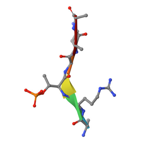
    Localization of the chromosomal passenger complex (CPC) at centromeres during early mitosis is essential for accurate chromosome segregation and is dependent on the phosphorylation of histone H3. We report the 2.7 Å resolution structure of the CPC subunit Survivin bound to the N-terminal tail of histone H3 carrying the Thr3 phosphorylation mark (Thr3ph). The BIR domain of Survivin recognizes the Ala1-Arg2-Thr3ph-Lys4 sequence, decoding the modification state and the free N terminus of histone H3 by a strategy similar to that used by PHD fingers. The structural analysis permitted the identification of putative Survivin-binding epitopes in other mitotic proteins, including human Shugoshin 1. Using biophysical and structural data, we show that a phospho-mimic N-terminal sequence such as that of hSgo1 (Ala1-Lys2-Glu3-Arg4) contains the specificity determinants to bind Survivin. Our findings suggest that the CPC engages in mutually exclusive interactions with other constituents of the mitotic machinery and a histone mark in chromatin.


  • Organizational Affiliation

    Department of Structural Cell Biology, Max-Planck-Institute of Biochemistry, Am Klopferspitz 18, D-82152 Martinsried, Germany. jeyaprak@biochem.mpg.de


Macromolecules
Find similar proteins by:  (by identity cutoff)  |  3D Structure
Entity ID: 1
MoleculeChains Sequence LengthOrganismDetailsImage
BACULOVIRAL IAP REPEAT-CONTAINING PROTEIN 5
A, B
142Homo sapiensMutation(s): 0 
UniProt & NIH Common Fund Data Resources
Find proteins for O15392 (Homo sapiens)
Explore O15392 
Go to UniProtKB:  O15392
PHAROS:  O15392
GTEx:  ENSG00000089685 
Entity Groups  
Sequence Clusters30% Identity50% Identity70% Identity90% Identity95% Identity100% Identity
UniProt GroupO15392
Sequence Annotations
Expand
  • Reference Sequence

Find similar proteins by:  Sequence   |   3D Structure  

Entity ID: 2
MoleculeChains Sequence LengthOrganismDetailsImage
HISTONE H3 PEPTIDE
C, D
6Homo sapiensMutation(s): 0 
UniProt & NIH Common Fund Data Resources
Find proteins for P68431 (Homo sapiens)
Explore P68431 
Go to UniProtKB:  P68431
PHAROS:  P68431
Entity Groups  
UniProt GroupP68431
Sequence Annotations
Expand
  • Reference Sequence
Small Molecules
Modified Residues  1 Unique
IDChains TypeFormula2D DiagramParent
TPO
Query on TPO
C, D
L-PEPTIDE LINKINGC4 H10 N O6 PTHR
Experimental Data & Validation

Experimental Data

  • Method: X-RAY DIFFRACTION
  • Resolution: 2.80 Å
  • R-Value Free: 0.268 
  • R-Value Work: 0.198 
  • R-Value Observed: 0.201 
  • Space Group: C 1 2 1
Unit Cell:
Length ( Å )Angle ( ˚ )
a = 114.365α = 90
b = 70.896β = 129.03
c = 81.465γ = 90
Software Package:
Software NamePurpose
PHENIXrefinement
XDSdata reduction
SCALAdata scaling
PHASERphasing

Structure Validation

View Full Validation Report


Entry History 

Deposition Data

Revision History  (Full details and data files)

  • Version 1.0: 2011-11-09
    Type: Initial release
  • Version 1.1: 2011-11-23
    Changes: Database references
  • Version 1.2: 2023-12-20
    Changes: Data collection, Database references, Derived calculations, Other, Refinement description