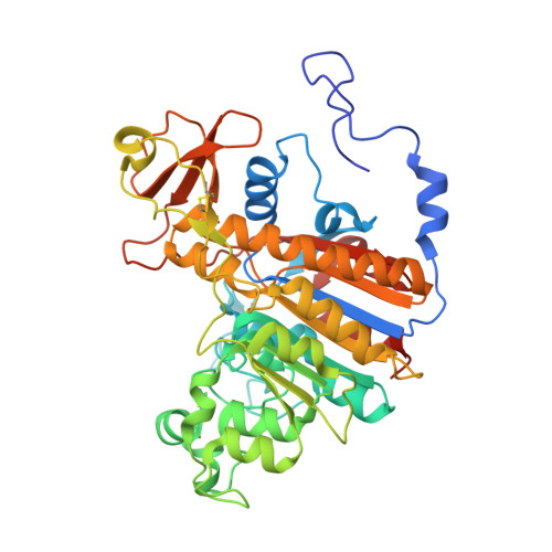
3TG0

E. coli alkaline phosphatase with bound inorganic phosphate


Experimental Data Snapshot

  • Method: X-RAY DIFFRACTION
  • Resolution: 1.20 Å
  • R-Value Free: 0.169 
  • R-Value Work: 0.148 
  • R-Value Observed: 0.149 

Starting Model: experimental
View more details

wwPDB Validation   3D Report Full Report


This is version 1.2 of the entry. See complete history


Literature

High-resolution analysis of Zn(2+) coordination in the alkaline phosphatase superfamily by EXAFS and x-ray crystallography.

Bobyr, E.Lassila, J.K.Wiersma-Koch, H.I.Fenn, T.D.Lee, J.J.Nikolic-Hughes, I.Hodgson, K.O.Rees, D.C.Hedman, B.Herschlag, D.

(2012) J Mol Biol 415: 102-117

  • DOI: https://doi.org/10.1016/j.jmb.2011.10.040
  • Primary Citation of Related Structures:  
    3TG0

  • PubMed Abstract: 

    Comparisons among evolutionarily related enzymes offer opportunities to reveal how structural differences produce different catalytic activities. Two structurally related enzymes, Escherichia coli alkaline phosphatase (AP) and Xanthomonas axonopodis nucleotide pyrophosphatase/phosphodiesterase (NPP), have nearly identical binuclear Zn(2+) catalytic centers but show tremendous differential specificity for hydrolysis of phosphate monoesters or phosphate diesters. To determine if there are differences in Zn(2+) coordination in the two enzymes that might contribute to catalytic specificity, we analyzed both x-ray absorption spectroscopic and x-ray crystallographic data. We report a 1.29-Å crystal structure of AP with bound phosphate, allowing evaluation of interactions at the AP metal site with high resolution. To make systematic comparisons between AP and NPP, we measured zinc extended x-ray absorption fine structure for AP and NPP in the free-enzyme forms, with AMP and inorganic phosphate ground-state analogs and with vanadate transition-state analogs. These studies yielded average zinc-ligand distances in AP and NPP free-enzyme forms and ground-state analog forms that were identical within error, suggesting little difference in metal ion coordination among these forms. Upon binding of vanadate to both enzymes, small increases in average metal-ligand distances were observed, consistent with an increased coordination number. Slightly longer increases were observed in NPP relative to AP, which could arise from subtle rearrangements of the active site or differences in the geometry of the bound vanadyl species. Overall, the results suggest that the binuclear Zn(2+) catalytic site remains very similar between AP and NPP during the course of a reaction cycle.


  • Organizational Affiliation

    Department of Chemistry, Stanford University, Stanford, CA 94305, USA.


Macromolecules
Find similar proteins by:  (by identity cutoff)  |  3D Structure
Entity ID: 1
MoleculeChains Sequence LengthOrganismDetailsImage
Alkaline phosphatase
A, B, C, D
449Escherichia coli K-12Mutation(s): 0 
Gene Names: phoAb0383JW0374
EC: 3.1.3.1
UniProt
Find proteins for P00634 (Escherichia coli (strain K12))
Explore P00634 
Go to UniProtKB:  P00634
Entity Groups  
Sequence Clusters30% Identity50% Identity70% Identity90% Identity95% Identity100% Identity
UniProt GroupP00634
Sequence Annotations
Expand
  • Reference Sequence
Small Molecules
Ligands 3 Unique
IDChains Name / Formula / InChI Key2D Diagram3D Interactions
PO4
Query on PO4

Download Ideal Coordinates CCD File 
H [auth A],
L [auth B],
P [auth C],
T [auth D]
PHOSPHATE ION
O4 P
NBIIXXVUZAFLBC-UHFFFAOYSA-K
ZN
Query on ZN

Download Ideal Coordinates CCD File 
E [auth A]
F [auth A]
I [auth B]
J [auth B]
M [auth C]
E [auth A],
F [auth A],
I [auth B],
J [auth B],
M [auth C],
N [auth C],
Q [auth D],
R [auth D]
ZINC ION
Zn
PTFCDOFLOPIGGS-UHFFFAOYSA-N
MG
Query on MG

Download Ideal Coordinates CCD File 
G [auth A],
K [auth B],
O [auth C],
S [auth D]
MAGNESIUM ION
Mg
JLVVSXFLKOJNIY-UHFFFAOYSA-N
Experimental Data & Validation

Experimental Data

  • Method: X-RAY DIFFRACTION
  • Resolution: 1.20 Å
  • R-Value Free: 0.169 
  • R-Value Work: 0.148 
  • R-Value Observed: 0.149 
  • Space Group: P 1 21 1
Unit Cell:
Length ( Å )Angle ( ˚ )
a = 65.963α = 90
b = 98.457β = 94.61
c = 152.146γ = 90
Software Package:
Software NamePurpose
Blu-Icedata collection
PHASERphasing
REFMACrefinement
DENZOdata reduction
SCALEPACKdata scaling

Structure Validation

View Full Validation Report


Entry History 

Deposition Data

Revision History  (Full details and data files)

  • Version 1.0: 2011-11-09
    Type: Initial release
  • Version 1.1: 2012-05-23
    Changes: Database references
  • Version 1.2: 2023-09-13
    Changes: Data collection, Database references, Derived calculations, Refinement description