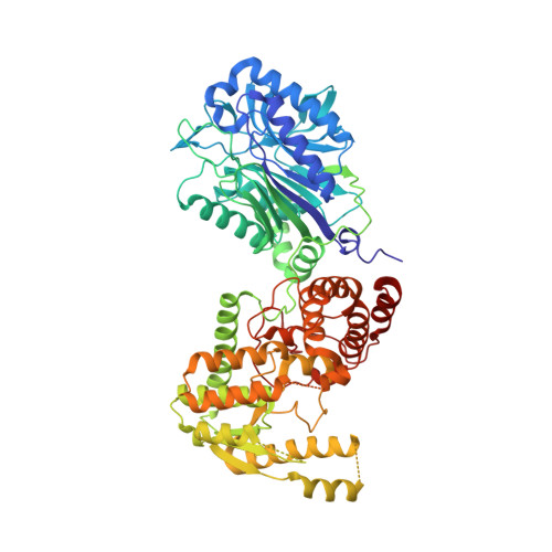
Regulation of the intersubunit ammonia tunnel in Mycobacterium tuberculosis glutamine-dependent NAD+ synthetase.
Chuenchor, W., Doukov, T.I., Resto, M., Chang, A., Gerratana, B.(2012) Biochem J 443: 417-426
- PubMed: 22280445
- DOI: https://doi.org/10.1042/BJ20112210
- Primary Citation of Related Structures:
3SDB, 3SEQ, 3SEZ, 3SYT, 3SZG - PubMed Abstract:
Glutamine-dependent NAD+ synthetase is an essential enzyme and a validated drug target in Mycobacterium tuberculosis (mtuNadE). It catalyses the ATP-dependent formation of NAD+ from NaAD+ (nicotinic acid-adenine dinucleotide) at the synthetase active site and glutamine hydrolysis at the glutaminase active site. An ammonia tunnel 40 Å (1 Å=0.1 nm) long allows transfer of ammonia from one active site to the other. The enzyme displays stringent kinetic synergism; however, its regulatory mechanism is unclear. In the present paper, we report the structures of the inactive glutaminase C176A variant in an apo form and in three synthetase-ligand complexes with substrates (NaAD+/ATP), substrate analogue {NaAD+/AMP-CPP (adenosine 5'-[α,β-methylene]triphosphate)} and intermediate analogues (NaAD+/AMP/PPi), as well as the structure of wild-type mtuNadE in a product complex (NAD+/AMP/PPi/glutamate). This series of structures provides snapshots of the ammonia tunnel during the catalytic cycle supported also by kinetics and mutagenesis studies. Three major constriction sites are observed in the tunnel: (i) at the entrance near the glutaminase active site; (ii) in the middle of the tunnel; and (iii) at the end near the synthetase active site. Variation in the number and radius of the tunnel constrictions is apparent in the crystal structures and is related to ligand binding at the synthetase domain. These results provide new insight into the regulation of ammonia transport in the intermolecular tunnel of mtuNadE.
- Department of Chemistry and Biochemistry, University of Maryland, College Park, MD 20742-2021, USA.
Organizational Affiliation:


