3REY | pdb_00003rey

Thermostabilised adenosine A2A receptor in complex with XAC


Experimental Data Snapshot

  • Method: X-RAY DIFFRACTION
  • Resolution: 3.31 Å
  • R-Value Free: 
    0.319 (Depositor), 0.360 (DCC) 
  • R-Value Work: 
    0.299 (Depositor), 0.310 (DCC) 
  • R-Value Observed: 
    0.300 (Depositor) 

Starting Model: experimental
View more details

wwPDB Validation 3D Report Full Report

Validation slider image for 3REY

Ligand Structure Quality Assessment 


This is version 1.3 of the entry. See complete history

Literature

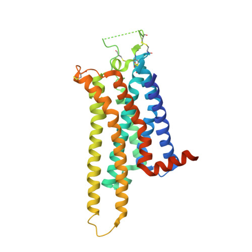
Structure of the adenosine A(2A) receptor in complex with ZM241385 and the xanthines XAC and caffeine

Dore, A.S.Robertson, N.Errey, J.C.Ng, I.Hollenstein, K.Tehan, B.Hurrell, E.Bennett, K.Congreve, M.Magnani, F.Tate, C.G.Weir, M.Marshall, F.H.

(2011) Structure 19: 1283-1293

  • DOI: https://doi.org/10.1016/j.str.2011.06.014
  • Primary Citation Related Structures: 
    3PWH, 3REY, 3RFM

  • PubMed Abstract: 

    Methylxanthines, including caffeine and theophylline, are among the most widely consumed stimulant drugs in the world. These effects are mediated primarily via blockade of adenosine receptors. Xanthine analogs with improved properties have been developed as potential treatments for diseases such as Parkinson's disease. Here we report the structures of a thermostabilized adenosine A(2A) receptor in complex with the xanthines xanthine amine congener and caffeine, as well as the A(2A) selective inverse agonist ZM241385. The receptor is crystallized in the inactive state conformation as defined by the presence of a salt bridge known as the ionic lock. The complete third intracellular loop, responsible for G protein coupling, is visible consisting of extended helices 5 and 6. The structures provide new insight into the features that define the ligand binding pocket of the adenosine receptor for ligands of diverse chemotypes as well as the cytoplasmic regions that interact with signal transduction proteins.


  • Organizational Affiliation
    • Heptares Therapeutics Ltd, BioPark, Welwyn Garden City, Herts, AL7 3AX, UK.

Macromolecule Content 

  • Total Structure Weight: 36.91 kDa 
  • Atom Count: 2,281 
  • Modeled Residue Count: 291 
  • Deposited Residue Count: 329 
  • Unique protein chains: 1

Macromolecules

Find similar proteins by:|  3D Structure
Entity ID: 1
MoleculeChains  Sequence LengthOrganismDetailsImage
Adenosine receptor A2a329Homo sapiensMutation(s): 8 
Gene Names: ADORA2A
Membrane Entity: Yes 
UniProt & NIH Common Fund Data Resources
Find proteins for P29274 (Homo sapiens)
Explore P29274 
Go to UniProtKB:  P29274
PHAROS:  P29274
GTEx:  ENSG00000128271 
Entity Groups
Sequence Clusters30% Identity50% Identity70% Identity90% Identity95% Identity100% Identity
UniProt GroupP29274
Sequence Annotations
Expand
Reference Sequence

Small Molecules

Ligands 1 Unique
IDChains Name / Formula / InChI Key2D Diagram3D Interactions
XAC

Query on XAC


Download:Ideal Coordinates CCD File
B [auth A]N-(2-aminoethyl)-2-[4-(2,6-dioxo-1,3-dipropyl-2,3,6,7-tetrahydro-1H-purin-8-yl)phenoxy]acetamide
C21 H28 N6 O4
FIQGIOAELHTLHM-UHFFFAOYSA-N

Experimental Data & Validation

Experimental Data

  • Method: X-RAY DIFFRACTION
  • Resolution: 3.31 Å
  • R-Value Free:  0.319 (Depositor), 0.360 (DCC) 
  • R-Value Work:  0.299 (Depositor), 0.310 (DCC) 
  • R-Value Observed: 0.300 (Depositor) 
Space Group: I 2 2 2
Unit Cell:
Length ( Å )Angle ( ˚ )
a = 111.865α = 90
b = 113.061β = 90
c = 126.802γ = 90
Software Package:
Software NamePurpose
ADSCdata collection
PHASERphasing
PHENIXrefinement
XDSdata reduction
SCALAdata scaling

Structure Validation

View Full Validation Report


Ligand Structure Quality Assessment 


Entry History 

Revision History  (Full details and data files)

  • Version 1.0: 2011-09-07
    Type: Initial release
  • Version 1.1: 2012-06-20
    Changes: Database references
  • Version 1.2: 2023-11-01
    Changes: Data collection, Database references, Derived calculations, Refinement description
  • Version 1.3: 2024-11-13
    Changes: Structure summary